42 thigh muscle chart
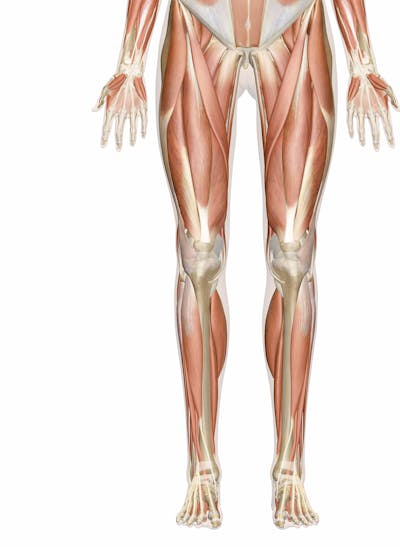
Muscles of the Thigh - Anterior - Medial - TeachMeAnatomy Muscles of the Thigh; Muscles in the Anterior Compartment of the Thigh. View Article. Muscles in the Medial Compartment of the Thigh. View Article. Muscles in the Posterior Compartment of the Thigh. View Article. Anatomy Video Lectures. START NOW FOR FREE. TeachMe Anatomy. Part of the TeachMe Series. Vivian Grisogono - ABOUT THE FRONT-THIGH MUSCLES Structure. The front-thigh consists of the quadriceps muscle group and the sartorius muscle, which lie over and around the thigh-bone (femur). There is also a small muscle called articularis genu, just above the front of the knee. Quadriceps means "four-headed", and the four parts which make up this muscle group are vastus lateralis, which ...
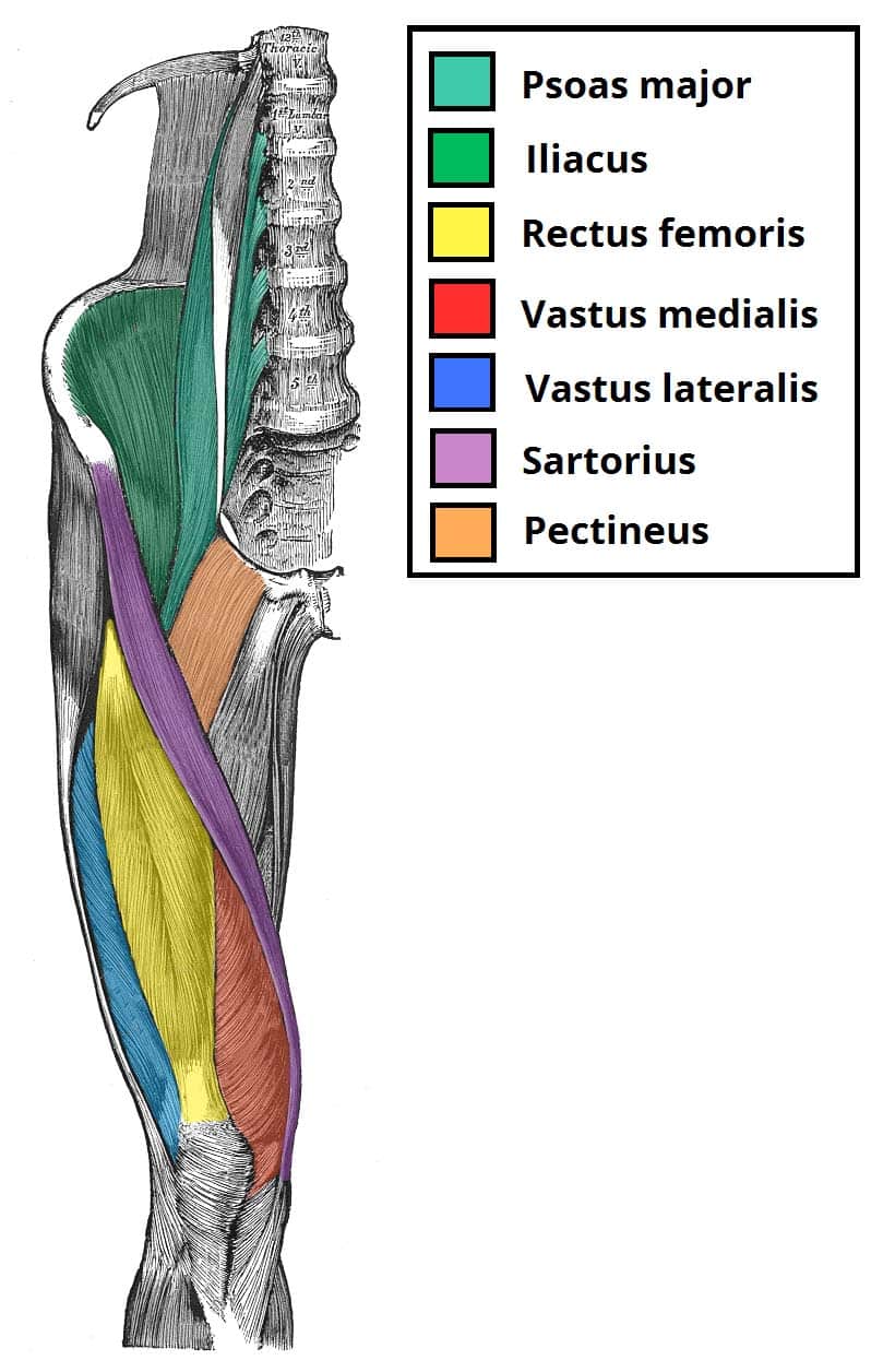
Muscles that act on the Anterior Thigh • Anatomy & Function Hip adduction is performed by the adductor magnus, adductor longus, adductor brevis and adductor minimus, as well as the pectineus muscle. The tensor fasciae latae muscle supports the gluteal muscles with the abduction of the thigh in the hip joint. It also stabilizes both the hip and knee joints. Learn anatomy faster and

Thigh muscle chart
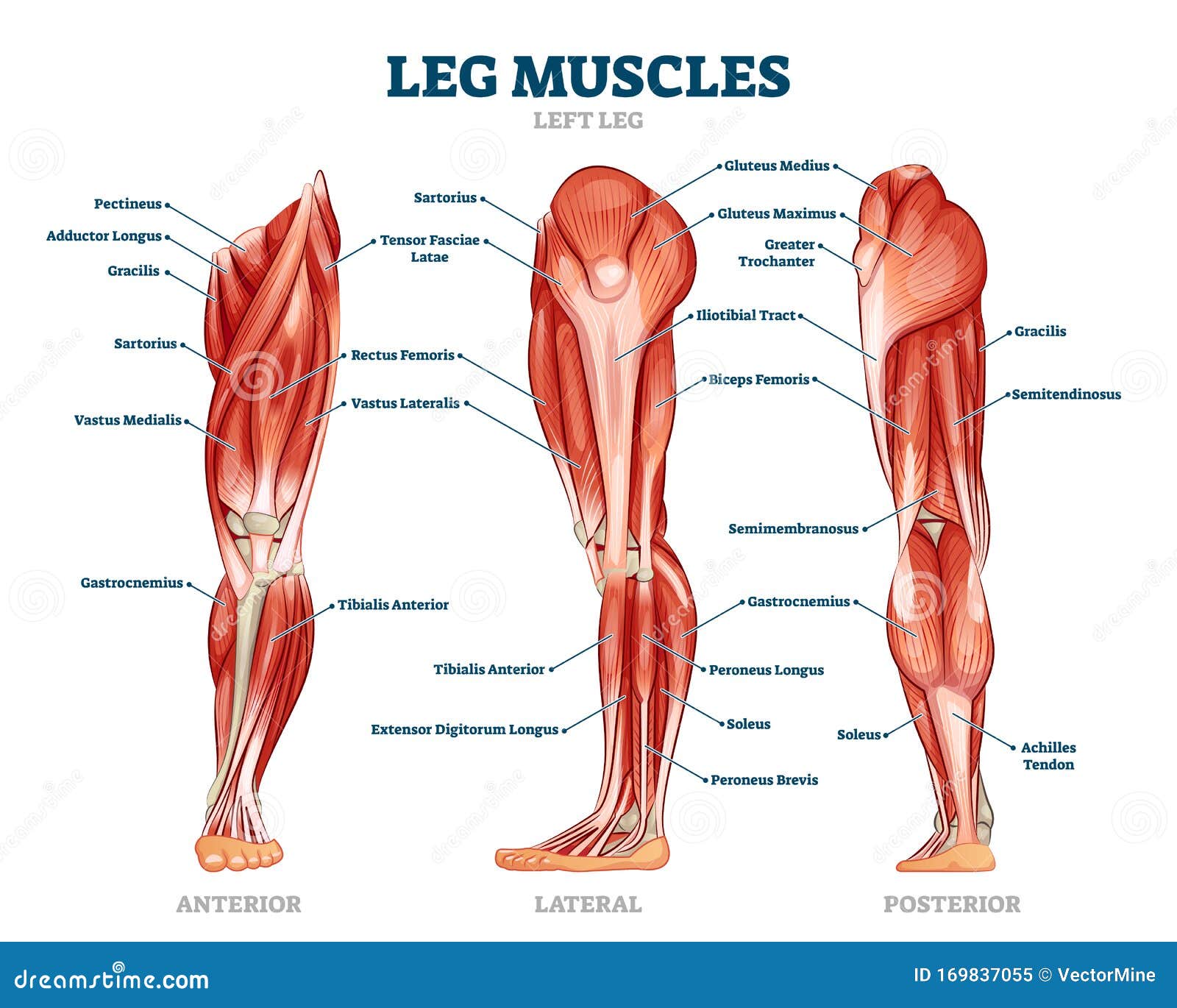
Leg Muscles: Anatomy and Function - Cleveland Clinic The muscles in your upper and lower legs work together to help you move, support your body's weight and allow you to have good posture. They enable you to do big movements, like running and jumping. They also help you with small movements, like wiggling your toes. Leg muscle strains are common, especially in the hamstrings, quads and groin. Thigh muscles Diagram | Quizlet Start studying Thigh muscles. Learn vocabulary, terms, and more with flashcards, games, and other study tools. Muscles of the Leg Laminated Anatomy Chart This chart is beautifully illustrated and offers the most comprehensive look at the muscles of the human leg available. Included are more than a dozen ...
Thigh muscle chart. Healthhype.com Muscle Pectinues Actions Adduction and flexion of the thigh Assists with medial rotation of the thigh Muscles Iliopsoas (psoas major, psoas minor, iliacus) Actions Flexion of the thigh at the hip joint Stabilizing the hip joint Muscle Sartorius Actions Abducts and flexes thigh at the hip joint Laterally rotates the the thigh at the hip joint Average Thigh Circumference and Size in Males and Females Additionally, you'll discover how to measure your thighs the right way so that you can get the most accurate measurement and see how your legs stack up. 14 inch thighs 17 inch thighs 18 inch thighs 19 inch thighs 20 inch thighs 21 inch thighs 22 inch thighs 23 inch thighs 24 inch thighs 25 inch thighs 26 inch thighs 27 inch thighs 28 inch thighs Leg Muscle Anatomy, Function, & Diagrams - Study.com The thigh of the leg has three major muscle groups to move the leg forward, backward, and towards the midline of the body. These muscles surround and control the movement of the femur, which is the... Muscle Charts of the Human Body — PT Direct For your reference value these charts show the major superficial and deep muscles of the human body. Superficial and deep anterior muscles of upper body Superficial and deep posterior muscles of upper body
human muscle system | Functions, Diagram, & Facts Aug 3, 2018 - human muscle system, the muscles of the human body that work the ... Human Anatomy and Physiology Diagrams: legs muscle diagram Leg Muscles ... Chart of Major Muscles on the Front of the Body with Labels A muscle of the medial thigh that originates on the pubis. It inserts onto the linea aspera of the femur. It adducts, flexes, and rotates the thigh medially. It is controlled by the obturator nerve. It pulls the leg toward the body's midline (i.e. adduction) Biceps brachii An upper arm muscle composed of 2 parts, a long head and a short head. leg muscle diagram deer hind quarter butcher whitetail leg. Untitled Document [bio.sunyorange.edu] bio.sunyorange.edu. sheep muscles thigh anatomy muscle goat leg system muscular human anterior posterior. Deer hind quarter butcher whitetail leg. Muscles evolution human sheep anatomy thigh posterior sunyorange updated2 bio edu. Diagram of my knee pain Muscles of the Leg Laminated Anatomy Chart - Pinterest The Muscles of the Leg anatomy chart shows in every possible view the way that the muscles and other pieces of the leg work together in motion and ...
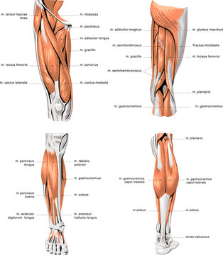
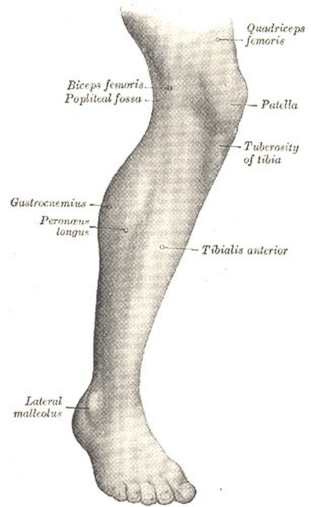
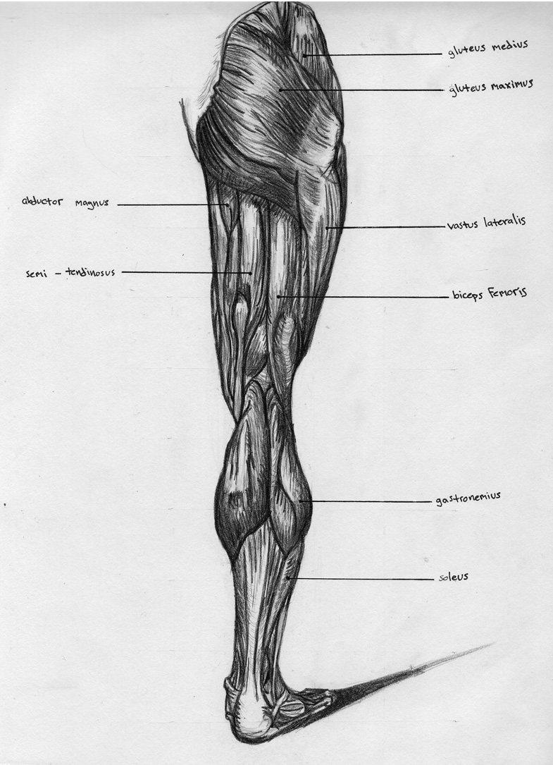
The Calf Muscle (Human Anatomy): Diagram, Function, Location The calf muscle, on the back of the lower leg, is actually made up of two muscles: The gastrocnemius is the larger calf muscle, forming the bulge visible beneath the skin. The gastrocnemius has two... Muscles of the Medial Thigh - TeachMeAnatomy The muscles in the medial compartment of the thigh are collectively known as the hip adductors. There are five muscles in this group; gracilis, obturator externus, adductor brevis, adductor longus and adductor magnus. All the medial thigh muscles are innervated by the obturator nerve, which arises from the lumbar plexus. Leg Muscle Diagram Pictures, Images and Stock Photos Human thigh muscle anatomy for the education. Muscular system legs Human muscular system of legs in back view. Gluteus medius, gluteus maximus gastrocnemius and other muscles. Pelvis, leg and hip bones skeleton poster. Bodybuilding and strong body vector illustration Cardiovascular System of the Leg and Foot 19 century medical illutrsation. Knee Muscles Anatomy - Pinterest Knee Muscles Anatomy, Anterior Leg Muscles, Anatomy Of The Knee, Upper Leg Muscles ... Arm Muscle Anatomy, Human Anatomy Chart, Physical Therapy School, ...
Muscles of the Leg Laminated Anatomy Chart - Pinterest The Muscles of the Leg anatomy chart shows in every possible view the way that the muscles and other pieces of the leg work together in motion and flexibility. This chart is beautifully illustrated and offers the most comprehensive look at the muscles of the human leg available. Included are more than a dozen illustrations like the vastus ...
Thigh Muscles: Anatomy, Function & Common Conditions Quadriceps include four large muscles located in the front of the thigh: vastus lateralis, vastus medialis, vastus intermedius, and rectus femoris. They start at the pelvis (hip bone) and femur (thigh bone) and extend down to the patella (kneecap) and tibia (shin bone). Sartorius muscle is a long, thin muscle — the longest in the human body.
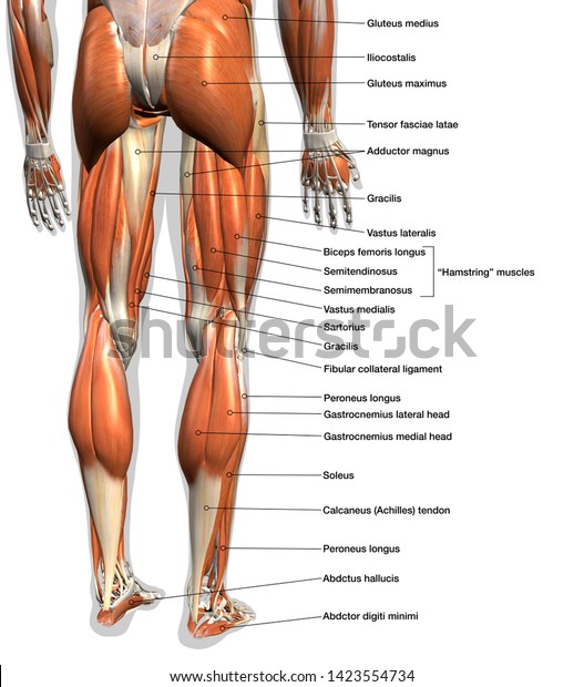
Leg: Anatomy and Function of Bones and Muscles, Plus Diagram Plantaris. This is a small muscle in the back of the lower leg. Like the gastrocnemius and soleus, it's involved in plantar flexion. Tibialis muscles. These muscles are found on the front and ...
legs muscle diagram - Pinterest The hand incorporates countless muscles, bones, tendons and ligaments into simple motion and this chart covers them all. There are over two dozen gorgeous and ...
Thigh Muscle Diagram - Pinterest The Muscles of the Leg anatomy chart shows in every possible view the way that the muscles and other pieces of the leg work together in motion and ...
leg muscle charts leg muscle charts gym workout chart - all-bodybuilding.com we have 8 Images about gym workout chart - all-bodybuilding.com like Chicken Muscle Anatomy Chart, Human Heart And Its Parts With Function Diagram Of Internal Parts Of and also gym workout chart - all-bodybuilding.com. Here you go: Gym Workout Chart - All-bodybuilding.com
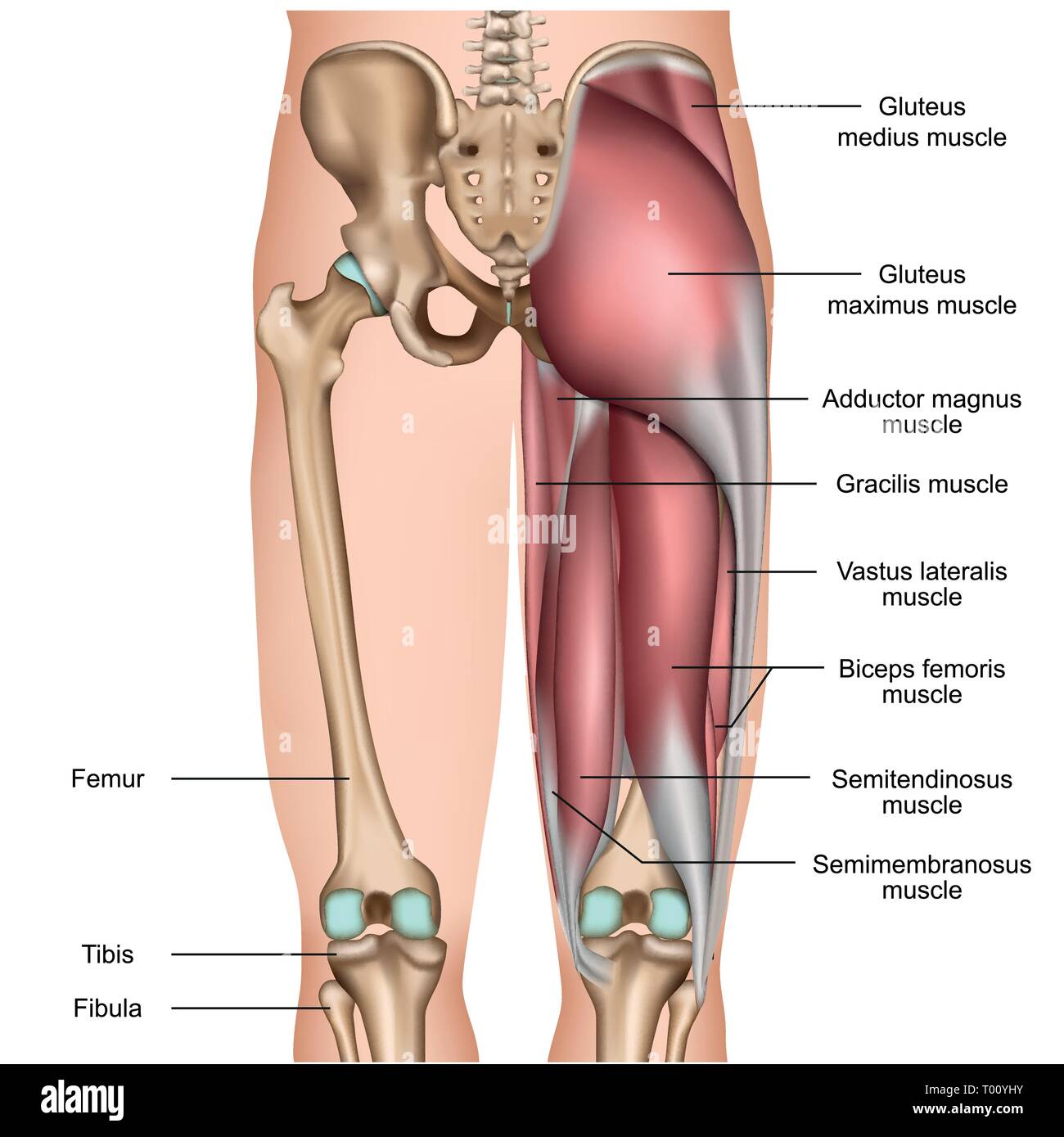
Thigh Anatomy, Diagram & Pictures | Body Maps - Healthline Muscles in the medial thigh help to bring the thigh toward the midline of the body and rotate it. These muscles are the adductor longus , adductor brevis , adductor magnus , gracilis, and the...
Leg Muscles Anatomy, Function & Diagram | Body Maps Gastrocnemius (calf muscle): One of the large muscles of the leg, it connects to the heel. It flexes and extends the foot, ankle, and knee. Soleus: This muscle extends from the back of the knee to ...
muscle diagram hip Hip anatomy thigh joint muscle posterior nerves muscles nerve supply kenhub diagram pelvic ligaments bones lumbosacral innervation knee. Investigation: Rat Dissection we have 6 Pics about Investigation: Rat Dissection like Leg Muscle Anatomy Chart | amulette, Hip and thigh: Bones, joints, muscles | Kenhub and also Muscle Bone Attachments.
Leg Muscles: Thigh and Calf Muscles, and Causes of Pain There are two main muscle groups in your upper leg. They include: Your quadriceps. This muscle group consists of four muscles in the front of your thigh which are among the strongest and largest...
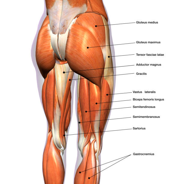
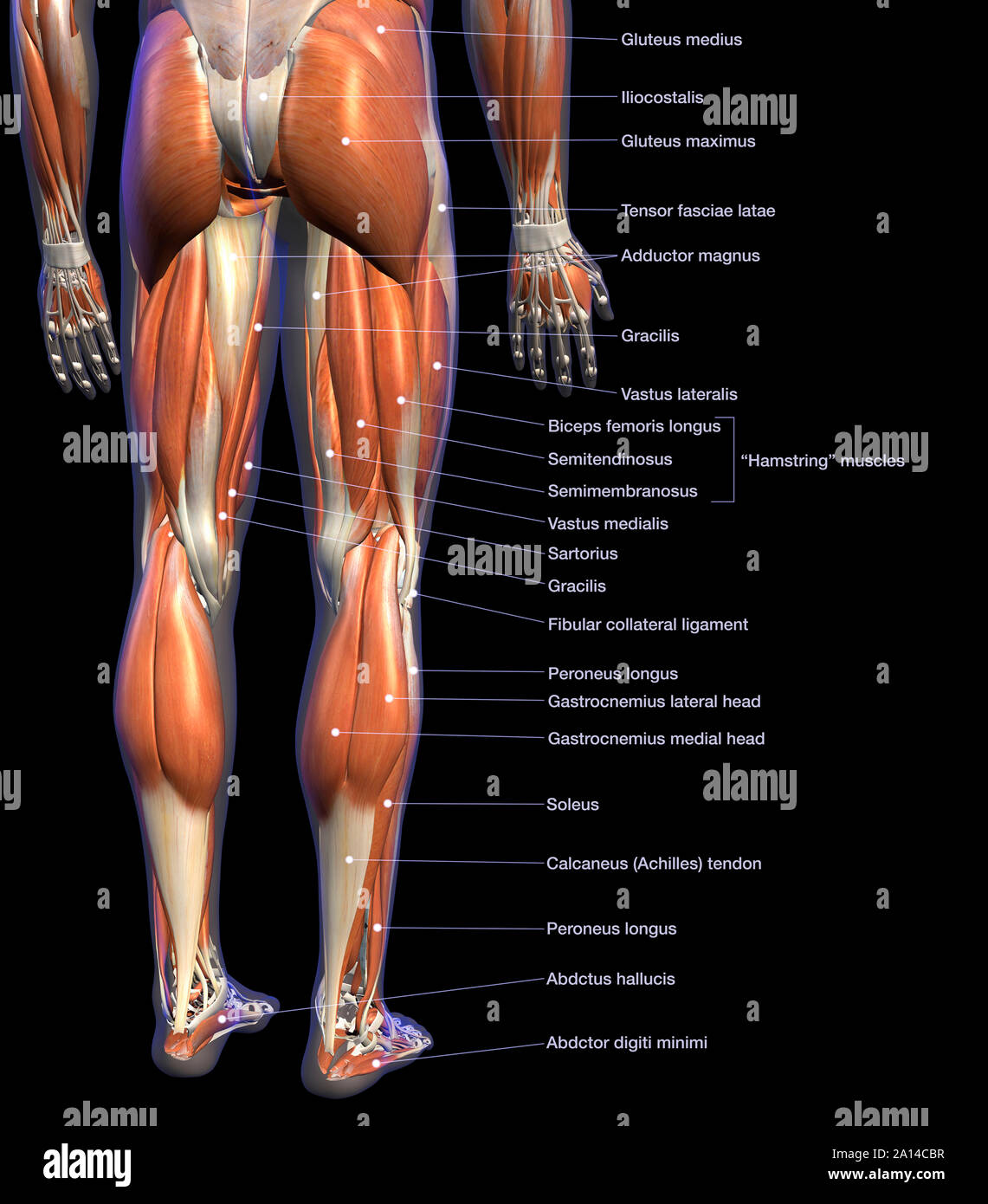
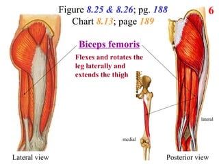
Hamstring Muscles: Location, Anatomy & Function The three hamstring muscles are: Biceps femoris, closest to the outside of your body. The function of this hamstring is to flex your knee, extend the thigh at your hip and rotate your lower leg from side-to-side when your knee is bent. Semimembranosus, closest to the middle of your body. This hamstring flexes your knee joint, extends your thigh ...
Hip and thigh muscles: Anatomy and functions | Kenhub The gluteal muscles can be divided into two main groups: Large and superficial muscles which mainly abduct and extend the thigh at the hip joint. These are the gluteus maximus, gluteus medius, gluteus minimus, and tensor fasciae latae. Small and deep muscles which mainly externally rotate the thigh at the hip joint and stabilize the pelvis.
Muscles of the hips and thighs - Course Hero Figure 9-8. The superficial muscles of the thigh. Figure 9-9. The quadriceps group of four muscles. The view on the left has the rectus femoris cut away to show the vastus intermedius which is below it. The sartorius muscle is a distinctively long and thin muscle that crosses the thigh diagonally. It is visible in Figure 9-8.
Thigh Muscle Diagram - Pinterest This chart is beautifully illustrated and offers the most comprehensive look at the muscles of the human leg available. Included are more than a dozen illustrations like the vastus lateralis, adductor brevis, rectus femoris, semi tendinosus and many, many more. Perfect for high level education or for patient consultations. This chart… Mark Munoz
thigh muscles diagram nerves leg muscles legs anatomy modernheal whatsapp. Lower Limb Nerve Anatomy Chart - Anterior - Chartex Ltd . lower anatomy limb anterior system nerve nerves nervous human major muscles chart upper charts femoral muscle body arteries spinal lumbar. Leg Nerves … | Physical Therapist Assistant, Anatomy, Physical Therapy
Lower Leg Muscle Diagram Blank Sketch Coloring Page Leg Muscles Diagram. Muscle Diagram. Lower Leg Muscles. Anatomy Practice. More information.... More like this. Head Muscles. Facial Muscles. Nursing Tips. Nursing Notes. Muscle Diagram. Anatomy Bones. Muscular System. Human Anatomy And Physiology. Muscle Anatomy. Muscle and anatomy are two words that are often heard when you are studying ...
Muscle anatomy reference charts: Free PDF download | Kenhub Lower limb (free PDF download) This muscle chart eBook covers the following regions: Inner hip & gluteal muscles. Anterior, medical and posterior thigh muscles. Anterior, lateral and posterior leg muscles. Dorsal and plantar foot muscles. This eBook contains high-quality illustrations and validated information about each muscle.
Muscles of the Leg Laminated Anatomy Chart This chart is beautifully illustrated and offers the most comprehensive look at the muscles of the human leg available. Included are more than a dozen ...
Thigh muscles Diagram | Quizlet Start studying Thigh muscles. Learn vocabulary, terms, and more with flashcards, games, and other study tools.
Leg Muscles: Anatomy and Function - Cleveland Clinic The muscles in your upper and lower legs work together to help you move, support your body's weight and allow you to have good posture. They enable you to do big movements, like running and jumping. They also help you with small movements, like wiggling your toes. Leg muscle strains are common, especially in the hamstrings, quads and groin.







:background_color(FFFFFF):format(jpeg)/images/library/14013/Hamstring_muscles.png)






![Muscular system legs - Stock Illustration [75914213] - PIXTA](https://en.pimg.jp/075/914/213/1/75914213.jpg)
Post a Comment for "42 thigh muscle chart"