43 microscope diagram to label
Label the microscope — Science Learning Hub 08/06/2018 · All microscopes share features in common. In this interactive, you can label the different parts of a microscope. Use this with the Microscope parts activity to help students identify and label the main parts of a microscope and then describe their functions.. Drag and drop the text labels onto the microscope diagram. If you want to redo an answer, click on the … Veterinary Anatomy » AnatomyLearner - The Place to Learn Veterinary ... 17/08/2022 17/08/2022 by anatomylearner. The dog shoulder anatomy consists primarily of a ball and socket joint between the glenoid cavity of the scapula and the head of a humerus. You will also see the infraspinatus, supraspinatus, biceps brachii, and teres major muscles in the dog shoulder structure. Again, you will find the capsular, medial ...
animal cell microscope picture - Theola Carranza Jun 15 2021 wga labeled cells were identified visually under the microscope captured ejected into pcr tubes containing 10 μl of lysis buffer and rnase inhibitor invitrogen 18080200 and flash frozen and stored at 80c until cdna synthesis. RF R98KEM Microbiological seamless pattern.
Microscope diagram to label
5 White Blood Cells Types and Their Functions - New Health Advisor Agranulocytes are free of visible grains under the microscope and include lymphocytes and monocytes. Together, they coordinate with one another to fight off things like cancer, cellular damage, and infectious diseases. Below, detailed information about each type will be discussed. 1. Neutrophils › 6-label-the-microscopeLabel the microscope — Science Learning Hub Jun 08, 2018 · All microscopes share features in common. In this interactive, you can label the different parts of a microscope. Use this with the Microscope parts activity to help students identify and label the main parts of a microscope and then describe their functions. Drag and drop the text labels onto the microscope diagram. If you want to redo an ... Nikon Instruments Inc. Nikon is a leader in microscope-based optical and imaging technologies for the life sciences and part of the Nikon Healthcare Business Division.
Microscope diagram to label. Electron microscope - Wikipedia An electron microscope is a microscope that uses a beam of accelerated electrons as a source of illumination. As the wavelength of an electron can be up to 100,000 times shorter than that of visible light photons, electron microscopes have a higher resolving power than light microscopes and can reveal the structure of smaller objects.. Electron microscopes use shaped magnetic … Diagram Label The Cellular Respiration - joi.hotelsalerno.sa.it cell cycle label - label a picture of the stages of mitosis, identify parts of the cell such as the centriole and spindle onion root tip lab - view real cells with a microscope, requires lab equipment and prepared slides the diagrams of the chloroplast (on the right below) and of the mitochondria (on the left below) onto your foldable write out … Ternary Phase Diagram - an overview | ScienceDirect Topics Ternary phase diagrams are used to represent all possible mixtures of three solvents [1]; they are described in Chapter 3.Here, we shall indicate how they should be used to minimize the solvent consumption. Figure 2.1 (top) shows the methanol–chloroform–water ternary phase diagram with the tie-lines in the biphasic domain. Five particular compositions are shown in the … Welcome to Virtual Urchin - University of Washington microscope measurement. microscope compare. specimen compare. development & embryology. fertilization lab. embryogenesis to hatching. analyzing gene function. ecology & environment. our acidifying ocean. predator & prey. surfing to settlement. basic biology. urchin anatomy. about us. teacher resources. useful links . Select Language: Welcome to the new …
ECLIPSE Ti2 Series | Inverted Microscopes | Nikon Microscope Products ... The ECLIPSE Ti2 inverted microscope delivers an unparalleled 25mm field of view (FOV) that revolutionizes the way you see. With this incredible FOV, the Ti2 maximizes the sensor area of large-format CMOS cameras without making compromises, and significantly improves data throughput. Epithelial Cell Organelles - types of epithelial cells stock image f031 ... Epithelial Cell Organelles - 18 images - the function and cell types of epithelial tissue, human epithelial cells stock video clip k003 6549 science photo, the cell the histology guide, euchromatin dr jastrows em atlas, Types of Muscle Contraction - TeachPE.com Types of Muscle Contraction - Isotonic, Concentric, Eccentric. Watch on. The three main types of muscle contraction during exercise are isotonic, isometric, and isokinetic. Also, we further categorise them into concentric and eccentric depending on whether the muscle shortens or lengthens when contracting. Meiosis Stages Quiz Questions And Answers - ProProfs Quiz Please take the quiz below, and it has some pictures to help you test out what you understand from the process of Meiosis as taught in biology class. Mark the answer! Have fun! Sorry if the pictures are small. There is a bonus problem. You'll know it when you see it. Questions and Answers. 1.
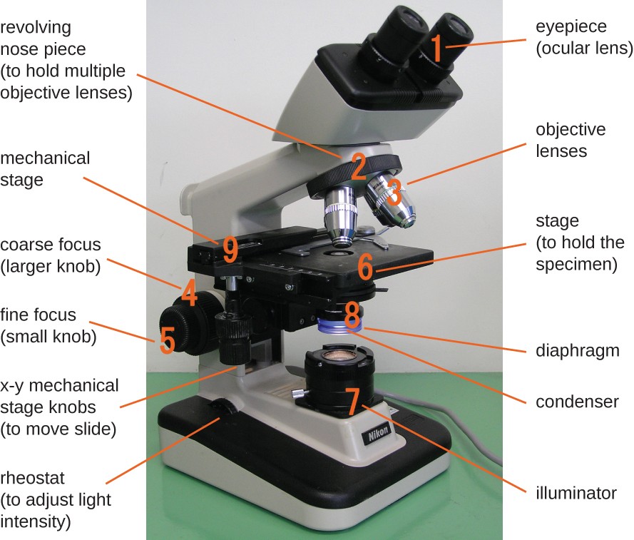
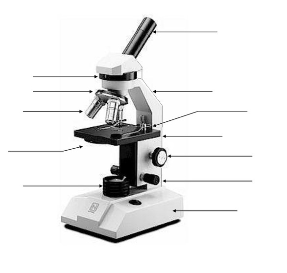
DNA sequencing - Wikipedia DNA sequencing is the process of determining the nucleic acid sequence - the order of nucleotides in DNA.It includes any method or technology that is used to determine the order of the four bases: adenine, guanine, cytosine, and thymine.The advent of rapid DNA sequencing methods has greatly accelerated biological and medical research and discovery. microbenotes.com › parts-of-a-microscopeParts of a microscope with functions and labeled diagram Apr 19, 2022 · Figure: Diagram of parts of a microscope. There are three structural parts of the microscope i.e. head, base, and arm. Head – This is also known as the body. It carries the optical parts in the upper part of the microscope. Base – It acts as microscopes support. It also carries microscopic illuminators. Parts of Stereo Microscope (Dissecting microscope) – labeled diagram ... Unlike a compound microscope that offers a flat image, stereo microscopes give the viewer a 3-dimensional image that you can see the texture of a larger specimen. [In this image] Examples of Stereo & Dissecting microscopes. Major microscope brands (Zeiss, Olympus, Nikon, Amscope, Omano, Leica …) all produce stereomicroscopes. Parts of a microscope with functions and labeled diagram 19/04/2022 · Figure: Diagram of parts of a microscope. There are three structural parts of the microscope i.e. head, base, and arm. Head – This is also known as the body. It carries the optical parts in the upper part of the microscope. Base – It acts as microscopes support. It also carries microscopic illuminators.
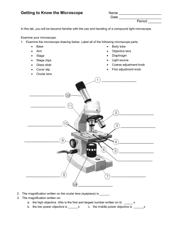
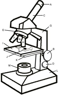
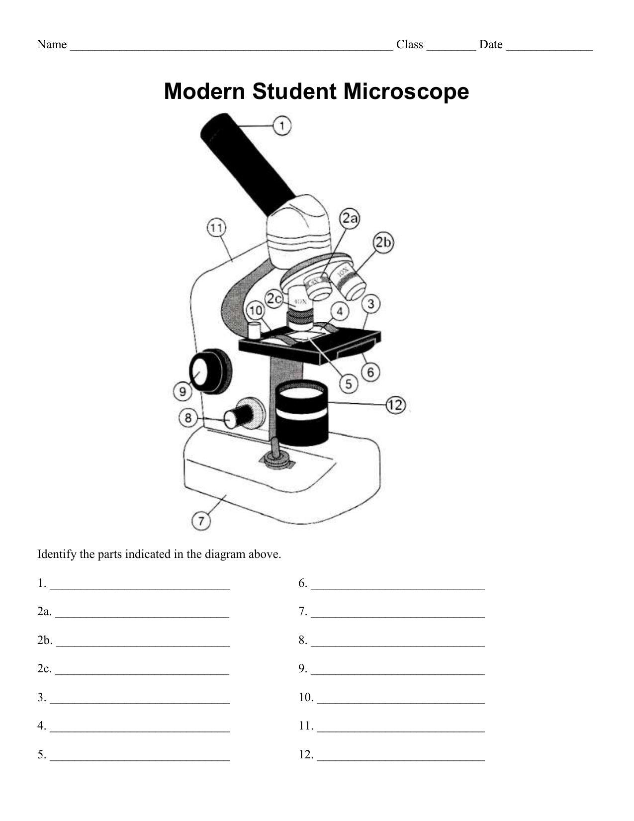
Getting to Know the Microscope | Manualzz
Metaphase - Genome.gov Metaphase is a stage during the process of cell division (mitosis or meiosis). Normally, individual chromosomes are spread out in the cell nucleus. During metaphase, the nucleus dissolves and the cell's chromosomes condense and move together, aligning in the center of the dividing cell. At this stage, the chromosomes are distinguishable when ...

Compound Microscope: Parts of Compound Microscope
researchtweet.com › microscope-parts-labeledMicroscope, Microscope Parts, Labeled Diagram, and Functions Jan 19, 2022 · The liquid sample comes next. To minimise evaporation and protect the microscope lens from sample exposure, a small square of clear glass or plastic (a coverslip) is placed on top of the liquid. 1. Collect a clean microscope slide and a coverslip (a thin piece of plastic covering). Fill the centre of the microscope slide with a drop or two of ...
Label microscope - Teaching resources
scheme work biology - Free KCPE Past Papers Introduction to light microscope. By the end of the lesson, the learner should be able to: Define a cell; Draw and label the light microscope; Description of a cell; Drawing and labeling the light microscope . Light microscope; Diagram of light microscope; Comprehensive secondary Biology students Bk. 1 page 17; Teachers bk. 1 pages 11-19; KLB ...

monocular labeling Diagram | Quizlet
Answers E Microscope Lab Letter - eluc.trattoria.napoli.it G Labeling Scientific Tools (Microscope) G F E D C B A 1) Illuminator 2) Stage 3) Eyepiece 4) Focus (Fine) 5) Lense 6) Focus (Course) 7) Base Determine which letter best matches each microscope piece .

Label microscope pt.1 Diagram | Quizlet
Spinal Cord Cross Section Explained (with Videos) Looking at a cross section of the spinal cord, you would see gray matter shaped like a butterfly surrounded by white matter. The gray matter is the core and ends up to be four projections that are known as horns. At the back are two dorsal horns and away from the back are two ventral horns.

Label the Microscope Diagram | Download Scientific Diagram
Fluorescence In Situ Hybridization (FISH) - Genome.gov The fluorescently labeled DNA finds its matching segment on one of the chromosomes, where it sticks. By looking at the chromosomes under a microscope, a researcher can find the region where the DNA is bound because of the fluorescent dye attached to it. This information thus reveals the location of that piece of DNA in the starting genome.

Microscope Maintenance Tips | Science supplies, Microscope ...
pE-300white | LED Microscope Illuminator - CoolLED Toxic mercury-based microscope illuminators are bad news for the environment, draw a lot of power and have a short lifetime. LED illumination systems are cleaner, have a long lifetime and use less power. Our pE-300 Series is also ACT label certified and is a natural choice for labs who want to play their part in helping the environment.
Microscope Diagram To Label - ClipArt Best - ClipArt Best ...
19 Types of Graphic Organizers for Effective Teaching and Learning Step 3: Write down the similarities in the bubbles that are common to both topics. Double Bubble Map Template (Click on the template to edit it online) 20. Venn diagram. Another graphic organizer that helps you visually represent a comparison of differences and similarities between two subjects, is the Venn diagram.

Microscope, Microscope Parts, Labeled Diagram, and Functions
Graphene - Wikipedia Graphene (/ ˈ ɡ r æ f iː n /) is an allotrope of carbon consisting of a single layer of atoms arranged in a two-dimensional honeycomb lattice nanostructure. The name is derived from "graphite" and the suffix -ene, reflecting the fact that the graphite allotrope of carbon contains numerous double bonds.. Each atom in a graphene sheet is connected to its three nearest neighbors by a strong ...
Instruments of Microscopy | Microbiology | | Course Hero
Mr. Jones's Science Class Earth, Moon, & Sun System (PPT.) Seasons Interactive (Online Activity) Moon Phases - Introductory Activity. Modeling the Phases of the Moon. Problems in Space (Online Activity) Lunar & Solar Eclipses - Webquest.

Parts of the Microscope with Labeling (also Free Printouts ...
The Elodea Under Microscope label the sketches to note the cell structures that you can identify complete the following questions: 1- describe the shape of the elodea leaf cells arm - portion of microscope that connects the body to the base base - a stable bottom for the microscope to stand upright (not labeled) iris diaphragm - allows light to settle onto slide ocular lens …
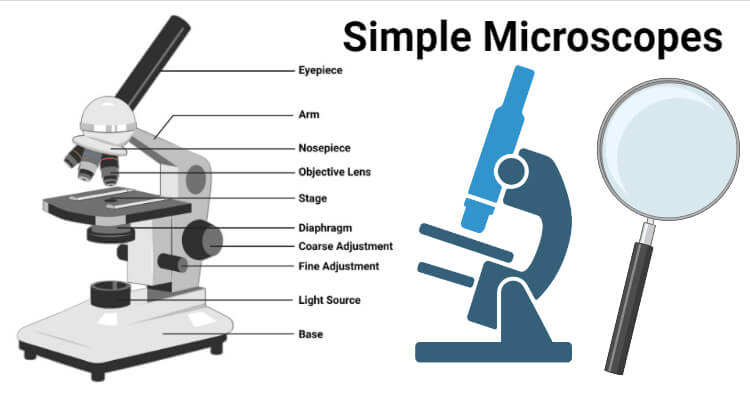
Simple Microscope- Definition, Principle, Magnification ...
Sternum: Anatomy, parts, pain and diagram | Kenhub The sternum is the bone that lies in the anterior midline of our thorax. It forms part of the rib cage and the anterior-most part of the thorax. Its functions are to protect the thoracic organs from trauma and also form the bony attachment for various muscles. It is also the center around which the superior 10 ribs directly or indirectly attached.
Microscope labeling, modern and classical types
Skeletal Muscle Structure Explained In Simple Terms - TeachPE.com The structure of skeletal muscle. In very simple terms, each muscle comprises bundles of muscles fibres which are made of bundles of myofibrils. Myofibrils divide along their length into Sarcomeres. Connective tissue runs through the muscles surrounding the various elements. Lets start at the outside and work inwards.

Microscope Labeling Diagram | Quizlet
en.wikipedia.org › wiki › Fluorescence_microscopeFluorescence microscope - Wikipedia A fluorescence microscope is an optical microscope ... design shown in the diagram. ... binding of an antibody to its antigen in order to label specific proteins or ...

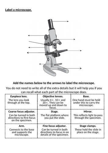
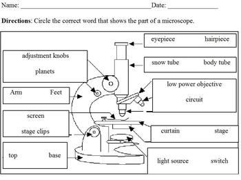
Label a Microscope Worksheet
Light Microscope (Theory) - Amrita Vishwa Vidyapeetham Microscope is an optical instrument that uses lens or combination of lens to produce magnified images that are too small to seen by unaided eye. Microscope provides the enlarged view that helps in examining and analyzing the image.

Draw a well labelled diagram of a microscope. - Brainly.in
DP Biology: Calculating Magnification and Size Click the eye icon to reveal a step by step calculation of specimen size using the scale bar. Activity 3 Calculating the size of a specimen using the magnification of the image Sometimes there is no scale bar but the magnification of the image is given. The image below shows three steps to calculate image size using the magnification.

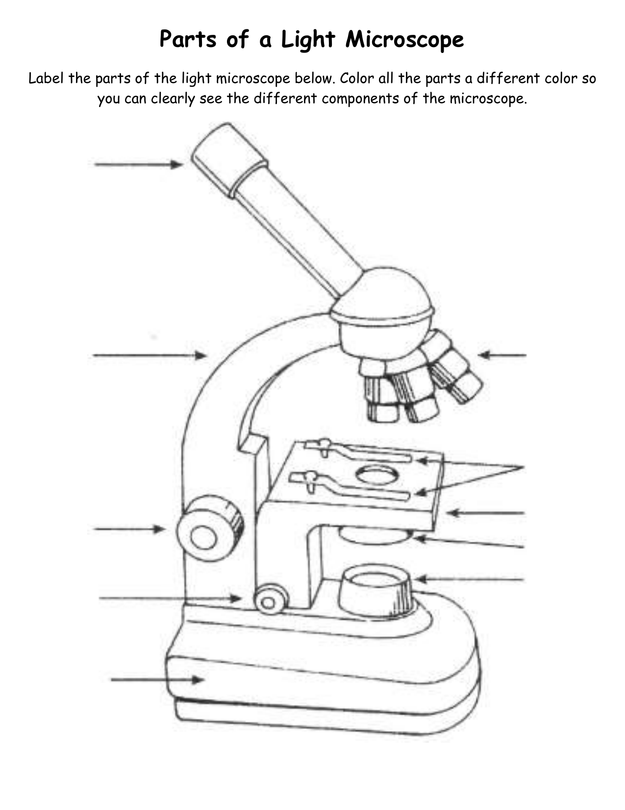
Parts of a Light Microscope Activity | Labeling Task
N-STORM | Super-Resolution Microscopes | Nikon Microscope Products ... N-STORM takes advantage of Nikon's powerful Ti2-E inverted microscope and applies high-accuracy, multi-color localization and reconstruction in three dimensions (xyz) to enable super-resolution imaging at tenfold the resolution of conventional light microscopes (up to approximately 20 nm in xy). This powerful technology enables the ...

Biology - labeling a compound microscope Diagram | Quizlet
The Obsessive Pleasures of Mechanical-Keyboard Tinkerers By David Owen. August 26, 2022. Illustration by Maria Chimishkyan. For months, Luke Bassett had been searching for a particular hard-to-find item, whose market value he estimated at a thousand ...

Microscope parts 3D learning for Android - APK Download
6M's | Cause & Effect Diagram | EdrawMax - Edrawsoft All-in-One Diagram Software Create more than 280 types of diagrams effortlessly Start diagramming with various templates and symbols easily Superior file compatibility: Import and export drawings to various file formats, such as Visio Cross-platform supported (Windows, Mac, Linux, Web) TRY IT FREE Security Verified | Switch to Mac >>

Microscope Diagram Labeled, Unlabeled and Blank | Parts of a ...
Microscope, Microscope Parts, Labeled Diagram, and Functions 19/01/2022 · Revolving Nosepiece or Turret: Turret is the part of the microscope that holds two or multiple objective lenses and helps to rotate objective lenses and also helps to easily change power. Objective Lenses: Three are 3 or 4 objective lenses on a microscope. The objective lenses almost always consist of 4x, 10x, 40x and 100x powers. The most common eyepiece …

Label Microscope Diagram | Microscope parts, Microscope ...
Large intestine: Anatomy, blood supply and innervation | Kenhub Anatomy. The large intestine is a 1 to 1.5 meter continuation of the ileum, extending from the ileocecal junction to the anus. Most of the large intestine is located inside the abdominal cavity, with the last portion residing within the pelvic cavity. Some parts of it are intraperitoneal while others are retroperitoneal .

Lable the microscope worksheet
Sketch Drawing Of A Microscope - Warehouse of Ideas Microscope View Represents The True/Exact Picture Of The Specimen In The Field Of View. Along with a drawing, a scientific sketch often includes labels and diagrams, questions and explanations. First of all sketch out a usual voluminous rectangle as in our example. Anatomy of a microscope microscope microscopic biology art.
Parts of a Microscope
Gram Stain Technique - Amrita Vishwa Vidyapeetham Wipe the glass slide with spirit and wave the slide over the Bunsen burner to remove any unwanted microorganisms in the slide. Label one side of the glass slide with 1. Your initials 2. The date While flaming the inoculation loop be sure that each segment of metal glows orange/red-hot before you move the next segment into the flame.

This is a common compound microscope. Label its parts from A ...
animal cell diagram labeled - Elouise Patel Labeled diagram of a typical animal cell Nucleus. Microscope cell staining is a technique used to improve the visibility of cells and cell parts under a microscope. It allows the orderly degradation and recycling of cellular components. The composition of cell wall is variable among the different groups of fungi.

Diagram of a Compound Microscope
depts.washington.edu › vurchinWelcome to Virtual Urchin - University of Washington Major update Apr 2021: All of the activities on the site are now mobile compatible !! Computers are still recommended, and tablets are preferable to phones: please read the Notes at the bottom of this page for details on the latest updates, mobile compatibility and general information about using this site.

Microscope labeled diagram
en.wikipedia.org › wiki › Electron_microscopeElectron microscope - Wikipedia An electron microscope is a microscope that uses a beam of accelerated electrons as a source of illumination. As the wavelength of an electron can be up to 100,000 times shorter than that of visible light photons , electron microscopes have a higher resolving power than light microscopes and can reveal the structure of smaller objects.

Compound Microscope Parts, Functions, and Labeled Diagram ...
rsscience.com › stereo-microscopeParts of Stereo Microscope (Dissecting microscope) – labeled ... Labeled part diagram of a stereo microscope Major structural parts of a stereo microscope. There are three major structural parts of a stereo microscope. The viewing Head includes the upper part of the microscope, which houses the most critical optical components, including the eyepiece, objective lens, and light source of the microscope.

Dissecting Stereo Microscope Parts and Functions
Fluorescence microscope - Wikipedia The majority of fluorescence microscopes, especially those used in the life sciences, are of the epifluorescence design shown in the diagram.Light of the excitation wavelength illuminates the specimen through the objective lens. The fluorescence emitted by the specimen is focused to the detector by the same objective that is used for the excitation which for greater resolution will …

Microscope Diagram - Free Printable Tests and Worksheets ...
Nikon Instruments Inc. Nikon is a leader in microscope-based optical and imaging technologies for the life sciences and part of the Nikon Healthcare Business Division.
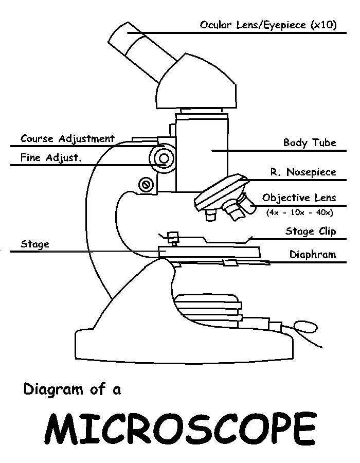
Diagram of a Microscope by ScienceDoodles on DeviantArt
› 6-label-the-microscopeLabel the microscope — Science Learning Hub Jun 08, 2018 · All microscopes share features in common. In this interactive, you can label the different parts of a microscope. Use this with the Microscope parts activity to help students identify and label the main parts of a microscope and then describe their functions. Drag and drop the text labels onto the microscope diagram. If you want to redo an ...
7Ac Microscope Labelling Worksheet | Teaching Resources
5 White Blood Cells Types and Their Functions - New Health Advisor Agranulocytes are free of visible grains under the microscope and include lymphocytes and monocytes. Together, they coordinate with one another to fight off things like cancer, cellular damage, and infectious diseases. Below, detailed information about each type will be discussed. 1. Neutrophils
Labeled Parts Of A Microscope - ClipArt Best

Free Microscope Drawing, Download Free Microscope Drawing png ...
Name Date Sci STANDARD MICROSCOPE DIAGRAM Label only the ...

Parts of a microscope with functions and labeled diagram

Labeling a Microscope Free Worksheet Pack
Modified Science Diagram; Label Parts of a Microscope; Special Education

Simple Microscope - Diagram (Parts labelled), Principle ...
Microscope, Microscope Parts, Labeled Diagram, and Functions

microscope drawing with label - Clip Art Library

Label the microscope — Science Learning Hub

Parts of the microscope activity
Free Microscope Drawing, Download Free Microscope Drawing png ...
Post a Comment for "43 microscope diagram to label"