45 labelled compound microscope
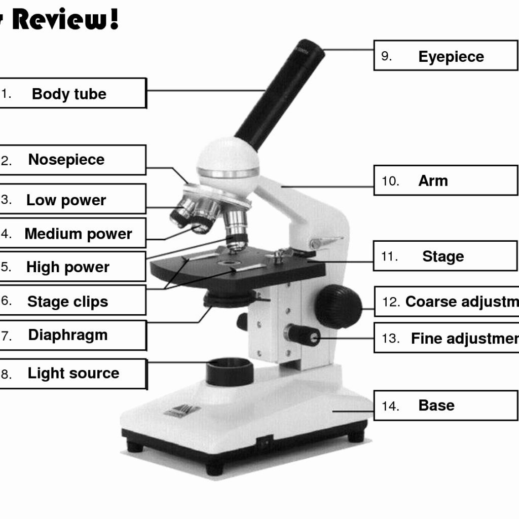
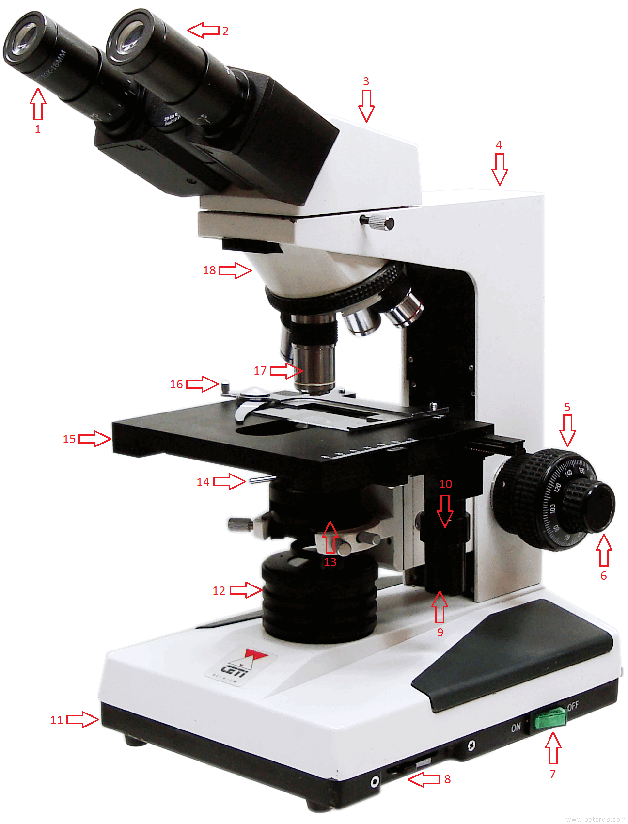
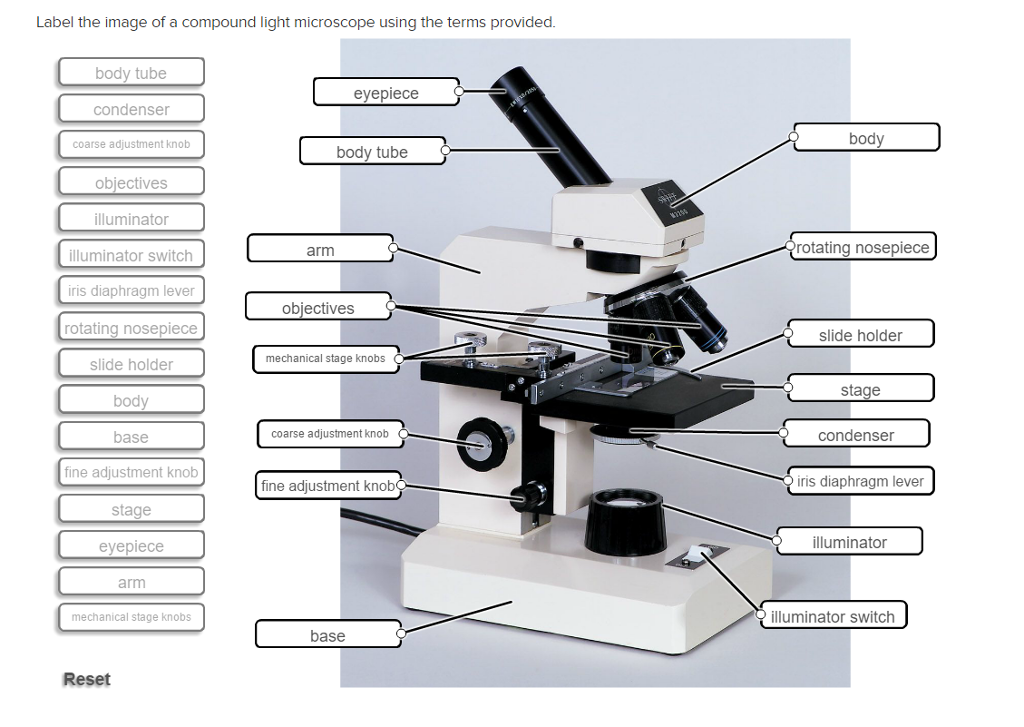
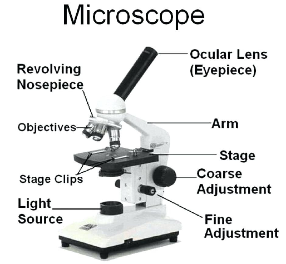
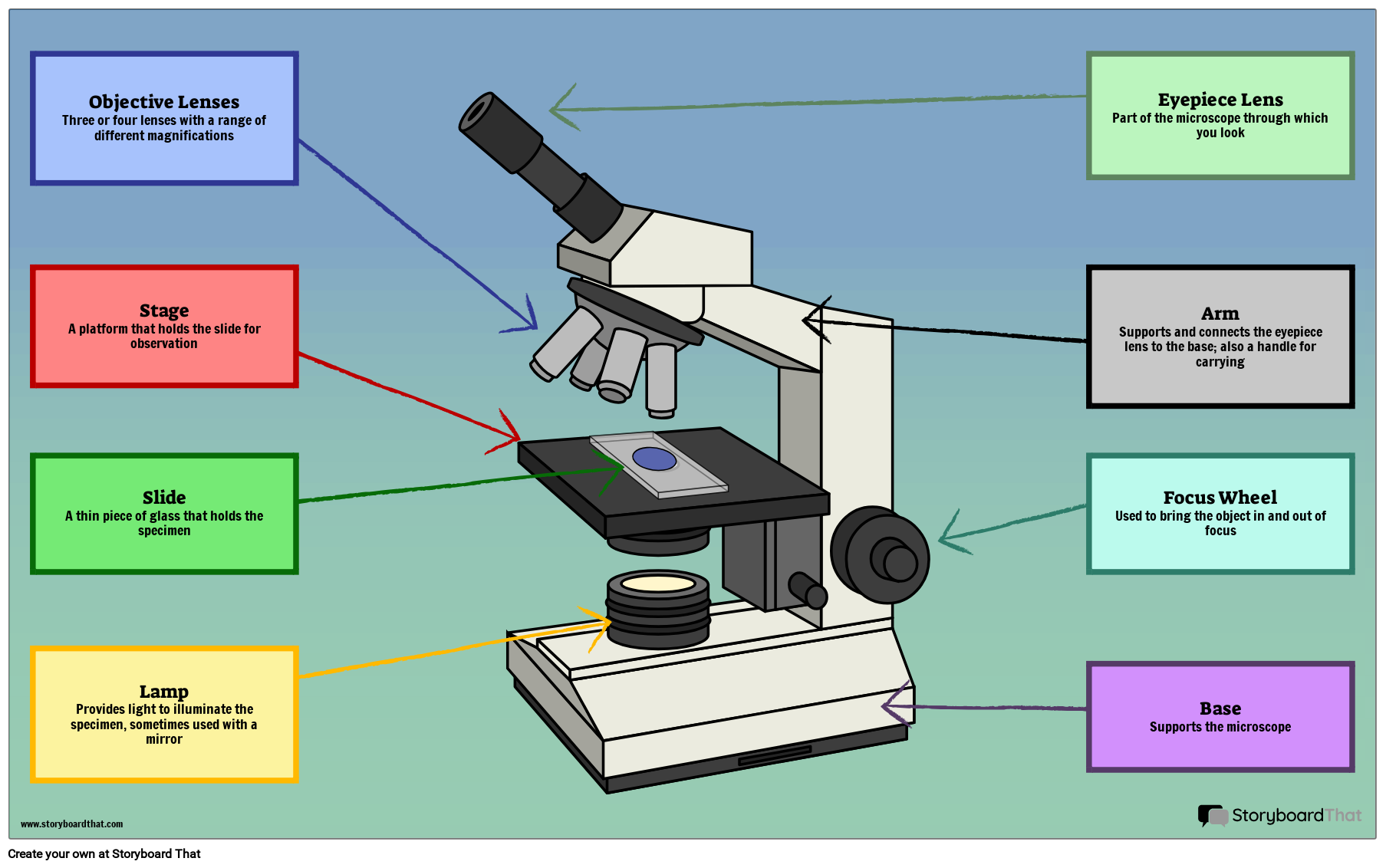
This is a common compound microscope. Label its parts ... This is a common compound microscope. Label its parts from A to J correctly. 1. Eye piece removed 2. Iris Diapharagm 3. Stage Plate 4. Eyepiece 5. Mirror 6. Compound Microscope - Diagram (Parts labelled), Principle and Uses A compound microscope: Is used to view samples that are not visible to the naked eye Uses two types of lenses - Objective and ocular lenses Has a higher level of magnification - Typically up to 2000x Is used in hospitals and forensic labs by scientists, biologists and researchers to study microorganisms
Parts of a Compound Microscope (And their Functions) - Scope Detective List of Microscope Parts and their Functions. 1. Ocular Tubes (Monocular, Binocular & Trinocular) The ocular tubes, are to tubes that lead from the head of the microscope out to your eyes. On the end of the ocular tubes are usually interchangeable eyepieces (commonly 10X and 20X) that increase magnification.

Labelled compound microscope
label parts of a compound microscope - TeachersPayTeachers Microscope Quiz by Quick Witted Owl Lesson Plans Made Easy 4 $1.99 PDF Students will need to label the parts of a compound light microscope and answer a 8 easy questions. A word bank is given at the bottom of the quiz. Compound Microscope Parts - Labeled Diagram and their Functions The term "compound" refers to the microscope having more than one lens. Basically, compound microscopes generate magnified images through an aligned pair of the objective lens and the ocular lens. In contrast, "simple microscopes" have only one convex lens and function more like glass magnifiers. A Study of the Microscope and its Functions With a Labeled Diagram ... The compound microscope uses light for illumination. Some compound microscopes make use of natural light, whereas others have an illuminator attached to the base. The specimen is placed on the stage and observed through different lenses of the microscope, which have varying magnification powers. Compound Microscope Parts and Functions
Labelled compound microscope. WO2022096473A1 - A positive electrode active material for … The present invention provides a positive electrode active oxide material for rechargeable batteries, comprising lithium, nickel, and at least one metal selected from the group comprising manganese and cobalt, whereby said positive electrode active material has a single-crystalline morphology and said surface layer further comprises aluminum and fluorine, wherein the atomic ratio of Al to a ... Draw a neat labelled diagram of a compound microscope ... The Optical Parts of Compound Microscope include: 1. Eyepiece lens or Ocular: At the top of the body tube, a lens is planted which is known as the eyepiece. On ... Binocular Microscope Anatomy - Parts and Functions with a Labeled ... Now, I will discuss the details anatomy of the light compound microscope with the labeled diagram. Why it is called binocular: because it has two ocular lenses or an eyepiece on the head that attaches to the objective lens, this ocular lens magnifies the image produced by the objective lens. Binocular microscope parts and functions Compound Microscope - Types, Parts, Diagram, Functions and Uses Compound microscope - It has two convex lenses. It is called a compound microscope because it compounds the light as it passes through the lenses to magnify. The image of the object being viewed is enlarged because of the lens near the object. An eyepiece, an additional lens, is where real magnification takes place.
Diagram of a Compound Microscope - Biology Discussion 1. It is noted first that which objective lens is in use on the microscope. 2. Stage micrometer is positioned in such a way that it is in the field of view. 3. The eyepiece is rotated so that the two scales, the eyepiece or ocular scale and the stage micrometer scale, are parallel. 4. Why Is The Light Microscope Called A Compound Microscope? These similarities create a bridge between the light and compound microscope, which sometimes confuses people. The light microscope is sometimes called a compound microscope because of its ability to use several lenses at a time, just like a compound microscope. Normal light microscopes cannot reach the highest magnifications because of their ... Compound Microscope- Definition, Labeled Diagram, Principle, Parts, Uses A compound microscope is of great use in pathology labs so as to identify diseases. Various crime cases are detected and solved by drawing out human cells and examining them under the microscope in forensic laboratories. The presence or absence of minerals and the presence of metals can be identified using compound microscopes. Label a Compound Microscope Diagram | Quizlet Start studying Label a Compound Microscope. Learn vocabulary, terms, and more with flashcards, games, and other study tools. ... Label this. Illuminator Switch. Sets found in the same folder. A&P II Ch. 24 Digestive Lab QUIZ. 10 terms. CWRN2016. Body planes Label. 9 terms. Hesi_Study.
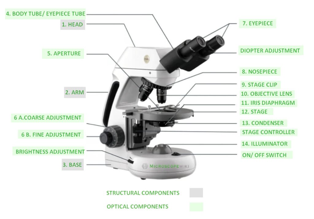
GitHub - Tirth27/Skin-Cancer-Classification-using-Deep ... EfficientNet used compound scaling (Figure 8), which uniformly scales the network's width, depth, and resolution. Among the different EfficientNet, EfficientNetB0 is the baseline network obtained by doing Neural Architecture Search (NAS). EfficientNetB1 to B7 is built upon the baseline network having a different value of compound scaling. Top 16 Techniques Used in Cell Biology (With Diagram) ADVERTISEMENTS: The following points highlight the top sixteen techniques used in cell biology. Some of the techniques are: 1. Immunofluorescence Microscopy 2. Ion-Exchange Chromatography 3. Affinity Chromatography 4. Partition and Adsorption Chromatography 5. Gel Filtration Chromatography 6. Radioactive Tracer Technique 7. Radioimmunoassay (RIA) 8. … Parts of a microscope with functions and labeled diagram - Microbe Notes There are three structural parts of the microscope i.e. head, base, and arm. Head - This is also known as the body. It carries the optical parts in the upper part of the microscope. Base - It acts as microscopes support. It also carries microscopic illuminators. Microscope Types (with labeled diagrams) and Functions A compound microscope: Is used to view samples that are not visible to the naked eye Uses two types of lenses - Objective and ocular lenses Has a higher level of magnification - Typically up to 2000x Is used in hospitals and forensic labs by scientists, biologists and researchers to study micro organisms Compound microscope labeled diagram
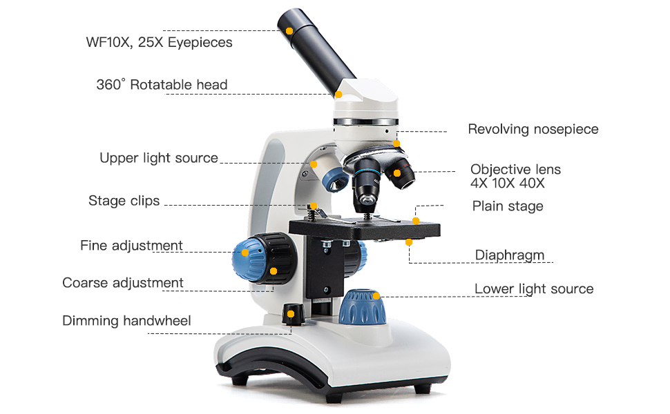
16 Parts of a Compound Microscope: Diagrams and Video Once you have an understanding of the parts of the microscope it will be much easier to navigate around and begin observing your specimen, which is the fun part! The 16 core parts of a compound microscope are: Head (Body) Arm. Base. Eyepiece. Eyepiece tube.
Parts of Stereo Microscope (Dissecting microscope) - labeled diagram ... Unlike a compound microscope that can only see a very thin specimen, stereo microscopes can be used for viewing almost anything you can fit under them. However, stereo microscopes offer lower magnification, typically 5x-50x, comparing to compound microscopes. Below is an example showing the difference between viewing with compound vs stereo ...
What is a Compound Microscope? - Study.com The body of the compound light microscope is the main part of the microscope, not to include the lights, focusing block, or stand of the microscope. The objective lenses and eyepiece are a part of ...
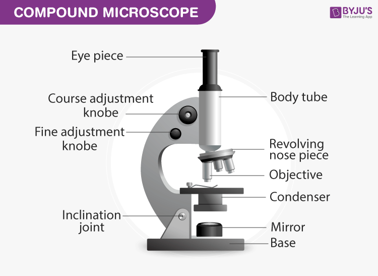
Compound Microscope: Parts of Compound Microscope - BYJUS (A) Mechanical Parts of a Compound Microscope 1. Foot or base It is a U-shaped structure and supports the entire weight of the compound microscope. 2. Pillar It is a vertical projection. This stands by resting on the base and supports the stage. 3. Arm The entire microscope is handled by a strong and curved structure known as the arm. 4. Stage
Microscope Parts and Functions First, the purpose of a microscope is to magnify a small object or to magnify the fine details of a larger object in order to examine minute specimens that cannot be seen by the naked eye. Here are the important compound microscope parts... Eyepiece: The lens the viewer looks through to see the specimen.
Compound Microscope Parts, Functions, and Labeled Diagram Compound Microscope Definitions for Labels. Eyepiece (ocular lens) with or without Pointer: The part that is looked through at the top of the compound microscope. Eyepieces typically have a magnification between 5x & 30x. Monocular or Binocular Head: Structural support that holds & connects the eyepieces to the objective lenses.
Compound Microscope Parts Head/Body houses the optical parts in the upper part of the microscope. Base of the microscope supports the microscope and houses the illuminator. Arm connects to the base and supports the microscope head. It is also used to carry the microscope. When carrying a compound microscope always take care to lift it by both the arm and base ...
Label the microscope — Science Learning Hub All microscopes share features in common. In this interactive, you can label the different parts of a microscope. Use this with the Microscope parts activity to help students identify and label the main parts of a microscope and then describe their functions. Drag and drop the text labels onto the microscope diagram.
Microbiological Examination of Foods: 7 Methods When examining foods, the possibility of detecting the presence of micro-organisms by looking at a sample directly under the microscope should not be missed. A small amount of material can be mounted and teased out in a drop of water on a slide, covered with a cover slip, and examined, first with a low magnification, and then with a x 45 objective.
What is a Compound Microscope? | Microscope World Blog A compound microscope is a high power (high magnification) microscope that uses a compound lens system. A compound microscope has multiple lenses: the objective lens (typically 4x, 10x, 40x or 100x) is compounded (multiplied) by the eyepiece lens (typically 10x) to obtain a high magnification of 40x, 100x, 400x and 1000x.
Labelled Diagram of Compound Microscope The below mentioned article provides a labelled diagram of compound microscope. Part # 1. The Stand: The stand is made up of a heavy foot which carries a curved inclinable limb or arm bearing the body tube. The foot is generally horse shoe-shaped structure (Fig. 2) which rests on table top or any other surface on which the microscope in kept.
A general design of caging-group-free photoactivatable ... - Nature 21.7.2022 · U2OS cells stably expressing a vimentin–HaloTag fusion construct 48 were labelled with compound 21 and imaged with a confocal microscope equipped with a subpicosecond pulsed laser (Fig. 3c).
A Study of the Microscope and its Functions ... - Pinterest May 21, 2019 - To better understand the structure and function of a microscope, we need to take a look at the labeled microscope diagrams of the compound ...
US10379038B2 - Measuring a size distribution of nucleic acid ... A process for measuring a size distribution of a plurality of nucleic acid molecules, the process comprising: labeling the nucleic acid molecules with a fluorescent dye comprising a plurality of fluorescent dye molecules to form labeled nucleic acid molecules, such that a number of fluorescent dyes molecules attached to each nucleic acid molecule is reliably proportional to the number of base ...
UK Technology Transfer - "UK Academia Technologies Available … The intercalate releases the pharmaceutically active compound in a controlled manner when subjected to low pH conditions that may prevail inside the stomach of a ... ‘click’ 18F-labelled octreotate PET imaging radiopharmaceutical that detects tumour lesions in patients with neuroendocrine ... Bright-field optical microscope converters, ...
Compound Microscope: Definition, Diagram, Parts, Uses, Working ... - BYJUS The compound microscope is mainly used for studying the structural details of cell, tissue, or sections of organs. The parts of a compound microscope can be classified into two: Non-optical parts Optical parts Non-optical parts Base The base is also known as the foot which is either U or horseshoe-shaped.
Effective uptake of submicrometre plastics by crop plants via ... - Nature 13.7.2020 · Two-week-old plants were exposed to 50 mg l –1 suspensions of PS beads labelled with NB and PS beads labelled with NBD-Cl for 10 d. The uptake of PS beads by wheat roots, stems and leaves was ...
Simple Microscope - Parts, Functions, Diagram and Labelling A compound microscope is also called a bright field microscope. It can provide magnification by up to 1,000 times. Stereo microscope/dissecting microscope - It can magnify objects by up to 300 times. It is used to visualize opaque objects that cannot be visualized using a compound microscope. Confocal microscope - It uses laser light to ...
Dextran - Wikipedia Dextran is a complex branched glucan (polysaccharide derived from the condensation of glucose), originally derived from wine. IUPAC defines dextrans as "Branched poly-α-d-glucosides of microbial origin having glycosidic bonds predominantly C-1 → C-6". Dextran chains are of varying lengths (from 3 to 2000 kilodaltons).. The polymer main chain consists of α-1,6 …
Labeling the Parts of the Microscope | Microscope World Resources Labeling the Parts of the Microscope This activity has been designed for use in homes and schools. Each microscope layout (both blank and the version with answers) are available as PDF downloads. You can view a more in-depth review of each part of the microscope here. Download the Label the Parts of the Microscope PDF printable version here.
Microscope Labeling - The Biology Corner Microscope Labeling. Shannan Muskopf May 31, 2018. This simple worksheet pairs with a lesson on the light microscope, where beginning biology students learn the parts of the light microscope and the steps needed to focus a slide under high power. The labeling worksheet could be used as a quiz or as part of direct instruction where students ...
Compound Microscope Labeled Diagram | Quizlet Compound Microscope Labeled + − Flashcards Learn Test Match Created by meganplocher734 Terms in this set (14) Eyepiece/Ocular lens Contains the ocular lens Body tube A hollow cylinder that holds the eyepiece. Arm Part that supports the microscope. Stage Supports the slide or specimen Coarse adjustment Knob
Parts of a Compound Microscope and Their Functions - NotesHippo Compound microscope mechanical parts (Microscope Diagram: 2) include base or foot, pillar, arm, inclination joint, stage, clips, diaphragm, body tube, nose piece, coarse adjustment knob and fine adjustment knob.. Base: It’s the horseshoe-shaped base structure of microscope.All of the other components of the compound microscope are supported by it. ...
A Study of the Microscope and its Functions With a Labeled Diagram ... The compound microscope uses light for illumination. Some compound microscopes make use of natural light, whereas others have an illuminator attached to the base. The specimen is placed on the stage and observed through different lenses of the microscope, which have varying magnification powers. Compound Microscope Parts and Functions
Compound Microscope Parts - Labeled Diagram and their Functions The term "compound" refers to the microscope having more than one lens. Basically, compound microscopes generate magnified images through an aligned pair of the objective lens and the ocular lens. In contrast, "simple microscopes" have only one convex lens and function more like glass magnifiers.
label parts of a compound microscope - TeachersPayTeachers Microscope Quiz by Quick Witted Owl Lesson Plans Made Easy 4 $1.99 PDF Students will need to label the parts of a compound light microscope and answer a 8 easy questions. A word bank is given at the bottom of the quiz.












Post a Comment for "45 labelled compound microscope"