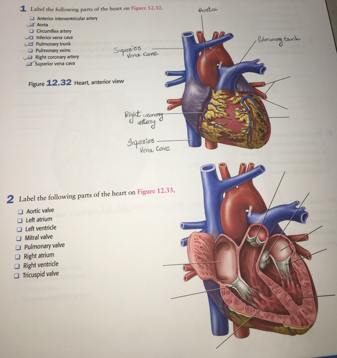
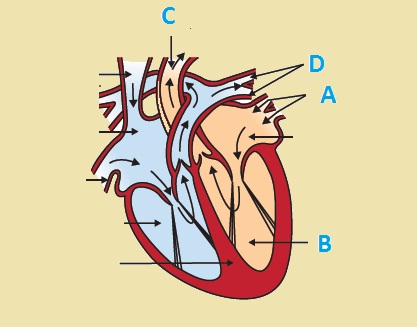
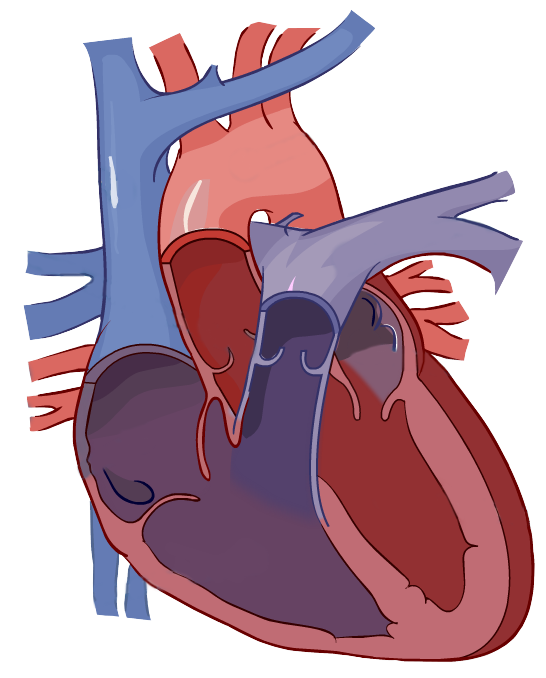
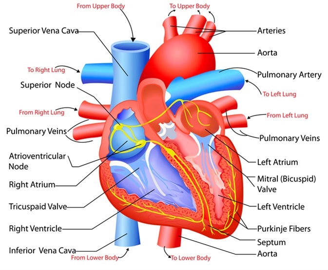
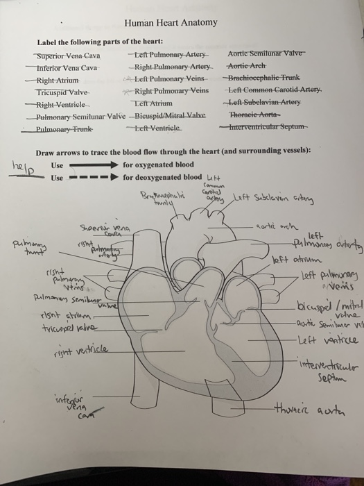
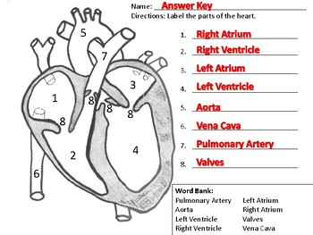
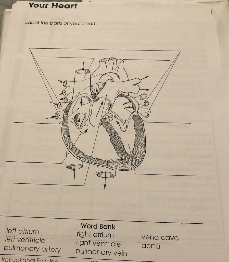
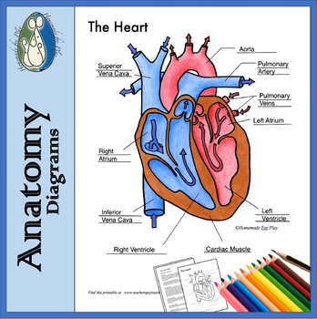
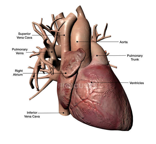
43 label the parts of a heart
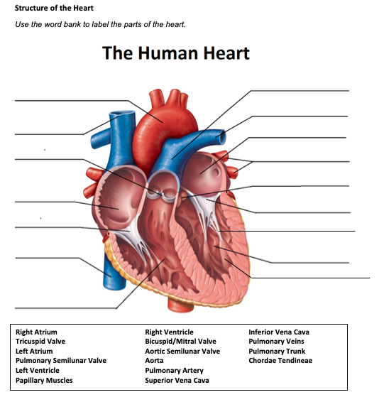
Human Heart Diagram Labeled - Science Trends The human heart usually weighs somewhere between 10 to 12 ounces in men and between 8 to 10 ounces in women, and in terms of size is roughly the size of the fist. The heart has four different chambers: the left and right ventricles and the left and right atriums. Solved Structure of the Heart Use the word bank to label the - Chegg 1. Human heart : It is a muscular organ that pumps the blood to the all parts of the body through cir … View the full answer Transcribed image text: Structure of the Heart Use the word bank to label the parts of the heart.
The Parts of the Heart and Their Functions | Life Persona 1 - The Epicardium Is the outermost layer of the heart. This serous membrane helps to lubricate and protect the exterior of the heart. 2 - The Myocardium Is the muscular layer of the heart. This is the thickest layer and is contracted involuntarily, allowing the heart to pump blood. 3 - The Endocardium Is the inner layer of the heart.
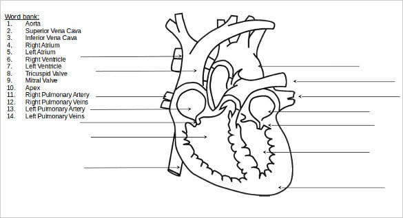
Label the parts of a heart
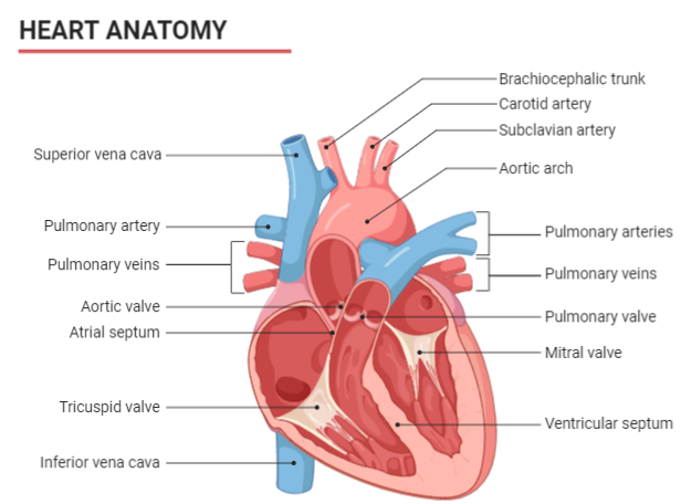
Label the Parts of the Heart Quiz - PurposeGames.com Label the Parts of the Heart — Quiz Information. This is an online quiz called Label the Parts of the Heart. There is a printable worksheet available for download here so you can take the quiz with pen and paper. From the quiz author. Utah Medical Anatomy and Physiology core Parts Of The Human Heart - Science Trends The parts of the human heart can be broken down into four chambers, muscular walls, vessels, and a conductive system. The two upper chambers are called the atria, with lower parts called ventricles. These all work together to make up the vital function of your heart. Everybody knows that the human heart is the essential organ in our bodies. Heart Diagram with Labels and Detailed Explanation - BYJUS The upper two chambers of the heart are called auricles. The lower two chambers of the heart are called ventricles. The heart wall is made up of three layers: The outer layer of the heart wall is called epicardium. The middle layer of the heart wall is called myocardium. The inner layer of the heart wall is called endocardium.
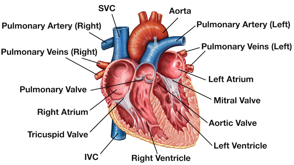
Label the parts of a heart. Label the Parts of the Heart Lessons, Worksheets and Activities Students Label the Parts of the Heart. Label the Parts of the Heart. Find the Resources You Need! Search . More Teaching Resources: Label the heart — Science Learning Hub Label the heart Interactive Add to collection In this interactive, you can label parts of the human heart. Drag and drop the text labels onto the boxes next to the diagram. Selecting or hovering over a box will highlight each area in the diagram. pulmonary vein semilunar valve right ventricle right atrium vena cava left atrium pulmonary artery A Labeled Diagram of the Human Heart You Really Need to See The human heart, comprises four chambers: right atrium, left atrium, right ventricle and left ventricle. The two upper chambers are called the left and the right atria, and the two lower chambers are known as the left and the right ventricles. The two atria and ventricles are separated from each other by a muscle wall called 'septum'. Labeling the parts of the heart. Flashcards | Quizlet Right Atrium Identify g Left Atrium Identify t Pulmonary Trunk Identify e Left Pulmonary Artery Identify r Right Pulmonary Artery Identify c Right Ventricle Identify j Left Ventricle Identify x Inferior Vena Cava Identify m Superior Vena Cava Identify b Acending Aorta Identify d Aortic Arch Identify p Apex Identify aa Left Pulmonary Veins
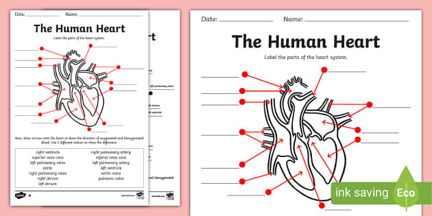
Label the Heart Diagram | Quizlet Right Ventricle ... Left Atrium ... Atrioventricular/Tricuspid Valve ... Atrioventricular/Mitral Valve ... Septum ... Right Atrium ... Semi-lunar Valves ... Left Pulmonary Veins ... Right Pulmonary Veins ... Left Pulmonary Arteries ... Right Pulmonary Arteries ... Left Ventricle ... Aorta ... Inferior Vena Cava ... Label parts of the heart - Liveworksheets Label parts of the heartDrag and drop the labels to the correct parts indicated on the heart diagram. ID: 832107. Language: English. School subject: Biology. Grade/level: GCSE. Age: 12-18. Main content: Label parts of the heart. Other contents: Add to my workbooks (516) Heart Anatomy: Labeled Diagram, Structures, Blood Flow ... - EZmed Chambers of the Heart Let's begin with the chambers of the heart. There are 4 chambers, labeled 1-4 on the diagram below. To help simplify things, we can convert the heart into a square. We will then divide that square into 4 different boxes which will represent the 4 chambers of the heart. Human Anatomy: Label the Heart Worksheets in 3 Differentiated Levels The Level 1 worksheet asks students to label the following parts of the heart: Aorta Vena Cava Right & Left Auricles Right & Left Ventricles The Level 2 worksheet asks students to label the above parts, plus the following: Superior and Inferior Vena Cava Aortic Arch Blood flowing to and from the body Blood flowing to and from the lungs
Structure of the Heart | SEER Training - National Cancer Institute Three layers of tissue form the heart wall. The outer layer of the heart wall is the epicardium, the middle layer is the myocardium, and the inner layer is the endocardium. Chambers of the Heart The internal cavity of the heart is divided into four chambers: Right atrium Right ventricle Left atrium Left ventricle Heart Labeling Quiz: How Much You Know About Heart Labeling? Here is a Heart labeling quiz for you. The human heart is a vital organ for every human. The more healthy your heart is, the longer the chances you have of surviving, so you better take care of it. Take the following quiz to know how much you know about your heart. Questions and Answers 1. What is #1? 2. What is #2? 3. What is #3? 4. What is #4? Label the parts of the dissected sheep heart. - Study.com Label the 5 parts of the heart conduction system. Draw a lymph node, and label its parts; Label where these parts are found. a. cerebral aqueduct b. fourth ventricle c. interventricular foramen d. lateral ventricles e. third ventricle; Draw a heart and label: a. both sets of valves and chambers of the heart b. the 2 major arteries exiting the heart Picture of the Heart - WebMD The heart is a muscular organ about the size of a fist, located just behind and slightly left of the breastbone. The heart pumps blood through the network of arteries and veins called the...
13 parts of the human heart (and its functions) - LORECENTRAL In general we can find the following parts of the heart . 1. Left atrium. One of the four main heart cavities in which blood is received and pumped . The left atrium is characterized by being connected to the pulmonary veins, from which it receives highly oxygenated blood and then sends it to the left ventricle. 2.
The Heart - Science Quiz - GeoGuessr The Heart - Science Quiz Aorta, Aortic valve, Left atrium, Left ventricle, Mitral valve, Pulmonary artery, Pulmonary valve, Pulmonary vein, Right atrium, Right ventricle, Septum, Superior vena cava, Tricuspid valve (13) Create custom quiz 0% | 0:06 | Click on: Right atrium > Sound On Review
Label Parts Of A Heart - Label The Heart Diagram Quizlet Label the following parts of the heart in the figure. 32 Label Parts Of The Heart Labels For Your Ideas from images.slideplayer.comCarries deoxygenated blood to the lungs · right . The human heart right atrium tricuspid valve right . Aortic valve left atrium left ventricle mitral valve pulmonary valve right atrium.
Label the Heart Quiz - PurposeGames.com There is a printable worksheet available for download here so you can take the quiz with pen and paper. From the quiz author Ummmmmmm . . . it's pretty self explanatory . . . you label the heart. Just remember one thing - you're looking at the heart like it's in someone else so right and left are switched around. Remaining 0 Correct 0 Wrong 0
The 18 parts of the human heart, and their functions The 18 parts of the human heart and how they work 1. Myocardium 2. Endocardium 3. Pericardium 4. Right Auricle 5. Right ventricle 6. Tricuspid valve 7. Pulmonary valve 8. Left Auricle 9. Left ventricle 10. Mitral valve 11. Aortic valve 12. Tendon cords 13. Papillary muscles 14. Sinus node 15. Atrioventricular node 16. Atrioventricular fascicule 17.
Labelling the heart — Science Learning Hub The heart is a muscular organ that pumps blood through the blood vessels of the circulatory system. Blood transports oxygen and nutrients to the body. It is also involved in the removal of metabolic wastes. In this activity, students use online and paper resources to identify and label the main parts of the heart.
Anatomy of a Human Heart - U of M Health Located between the lungs in the middle of the chest, the heart pumps blood through the network of arteries and veins known as the cardiovascular system. It pushes blood to the body's organs, tissues and cells. Blood delivers oxygen and nutrients to every cell and removes the carbon dioxide and other waste products made by those cells.
The Anatomy of the Heart, Its Structures, and Functions - ThoughtCo The heart is the organ that helps supply blood and oxygen to all parts of the body. It is divided by a partition (or septum) into two halves. The halves are, in turn, divided into four chambers. The heart is situated within the chest cavity and surrounded by a fluid-filled sac called the pericardium. This amazing muscle produces electrical ...
Diagram of Human Heart and Blood Circulation in It The wall of the heart has three different layers, such as the Myocardium, the Epicardium, and the Endocardium. Here's more about these three layers. Epicardium The outermost layer of your heart wall is called the epicardium, which is basically a very thin layer of serous membrane.
Human Heart - Anatomy, Functions and Facts about Heart - BYJUS Following are the main functions of the heart: One of the primary functions of the human heart is to pump blood throughout the body. Blood delivers oxygen, hormones, glucose and other components to various parts of the body, including the human heart. The heart also ensures that adequate blood pressure is maintained in the body.
Heart Diagram with Labels and Detailed Explanation - BYJUS The upper two chambers of the heart are called auricles. The lower two chambers of the heart are called ventricles. The heart wall is made up of three layers: The outer layer of the heart wall is called epicardium. The middle layer of the heart wall is called myocardium. The inner layer of the heart wall is called endocardium.
Parts Of The Human Heart - Science Trends The parts of the human heart can be broken down into four chambers, muscular walls, vessels, and a conductive system. The two upper chambers are called the atria, with lower parts called ventricles. These all work together to make up the vital function of your heart. Everybody knows that the human heart is the essential organ in our bodies.
Label the Parts of the Heart Quiz - PurposeGames.com Label the Parts of the Heart — Quiz Information. This is an online quiz called Label the Parts of the Heart. There is a printable worksheet available for download here so you can take the quiz with pen and paper. From the quiz author. Utah Medical Anatomy and Physiology core





















Post a Comment for "43 label the parts of a heart"