41 microscope diagram with labels
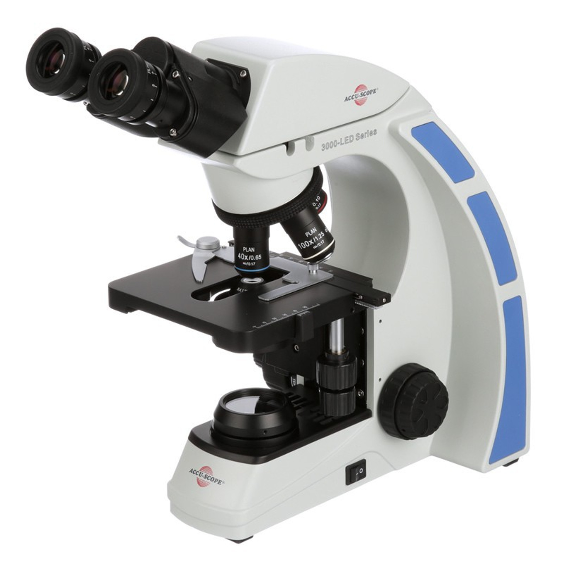
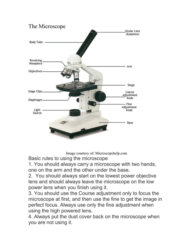
Microscope, Microscope Parts, Labeled Diagram, and Functions The Microscopes parts divided into three different structural parts Head, Base, and Arms. Head/Body: It contain the optical parts in the upper part of the microscope. Arm: It supports the tube and connects it to the base. Base: The bottom of the microscope, used for support. Optical Components of Microscope Microscope: Parts Of A Microscope With Functions And Labeled Diagram. The microscope has three basic components: the head, the base, and the arm. Head:Occasionally, the head is considered the body. It holds the optical components of the upper part of the microscope. Base:The microscope's base provides great support. It is also equipped with miniature illuminators.
Parts of Stereo Microscope (Dissecting microscope) - labeled diagram ... Labeled part diagram of a stereo microscope Major structural parts of a stereo microscope Optical components of a stereo microscope - definition and function Eyepieces Eyepiece tube Diopter adjustment ring Interpupillary Adjustment Objective Lenses Barlow lens Adjustment Knobs Light sources Stage plate Stage chips
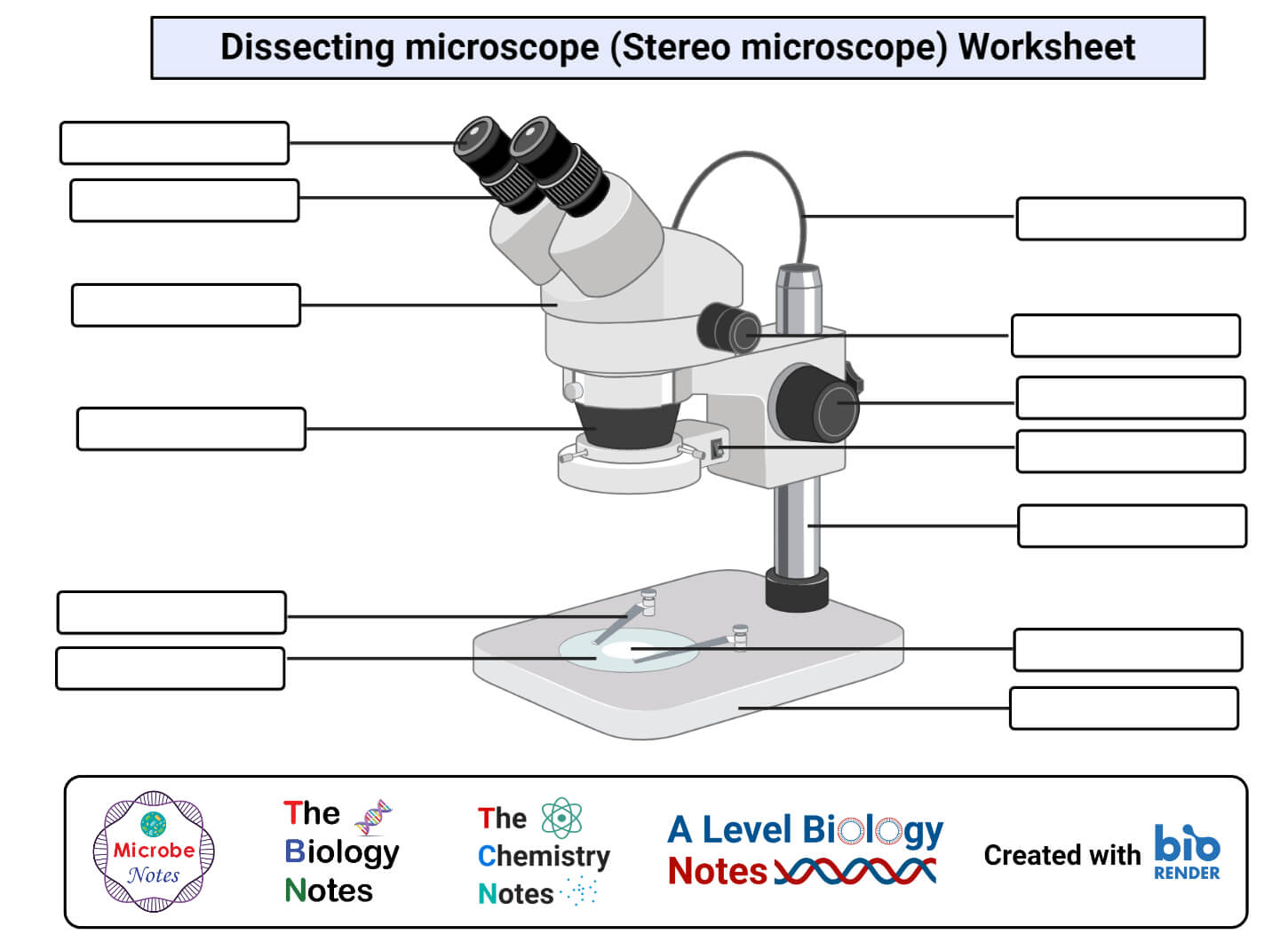
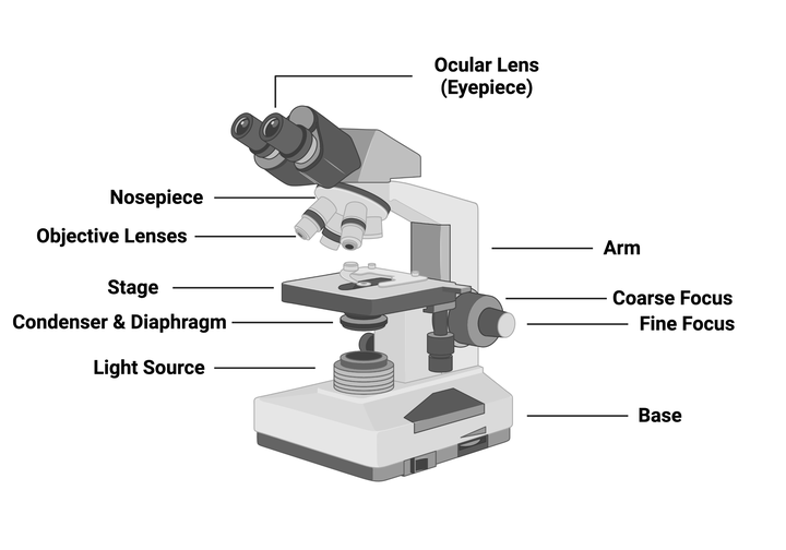
Microscope diagram with labels
Parts of the Microscope with Labeling (also Free Printouts) A microscope is one of the invaluable tools in the laboratory setting. It is used to observe things that cannot be seen by the naked eye. Table of Contents 1. Eyepiece 2. Body tube/Head 3. Turret/Nose piece 4. Objective lenses 5. Knobs (fine and coarse) 6. Stage and stage clips 7. Aperture 9. Condenser 10. Condenser focus knob 11. Iris diaphragm Microscope Parts, Types & Diagram | What is a Microscope? Microscope Diagram There are many illustrations available for the diagram of a light microscope. The essential parts include the head, base, arms, lenses, and lights. In diagrams, one... Parts of the Microscope (Labeled Diagrams) Simple microscope labelled diagram Image created with Biorender Tube/Body Tube It serves as the connector between the eyepiece/ocular and objective lenses. Objective lenses The lenses have varying magnifying power, which typically consists of 10x, 40x, and 100x.
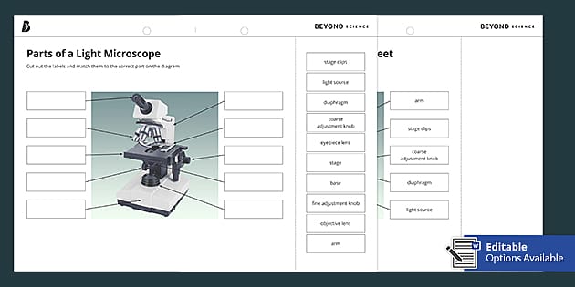
Microscope diagram with labels. Parts of a microscope with functions and labeled diagram - Microbe Notes Parts of a microscope with functions and labeled diagram September 17, 2022 by Faith Mokobi Having been constructed in the 16th Century, Microscopes have revolutionalized science with their ability to magnify small objects such as microbial cells, producing images with definitive structures that are identifiable and characterizable. Label a microscope - Labelled diagram Eyepiece (x10), Arm, Diaphragm, Coarse adjustment, Fine adjustment, Base, Light, Stage, Clips, Objective x4, Objective x10, Objective x 40. Microscope Labeling Diagram | Quizlet Contains the ocular lens and magnifies the image produced by the objective lenses. Location Term Coarse Focus Knob Definition Moves the stage large distances to roughly focus the image. Location Term Fine Focus Knob Definition Moves the stage tiny distances to slightly adjust and fine-tune the image focus. Location Term Arm Definition 16 Essential Microscope Parts: Names, Functions & Labeled Diagram Microscope Parts Labeled Diagram The principle of the Microscope gives you an exact reason to use it. It works on the 3 principles. Magnification Resolving Power Numerical Aperture. Parts of Microscope Head Base Arm Eyepiece Lens Eyepiece Tube Objective Lenses Nose Piece Adjustment Knobs Stage Aperture Microscopic Illuminator Condenser Lens
Microscope Diagram - Label Diagram | Quizlet Start studying Microscope Diagram - Label. Learn vocabulary, terms, and more with flashcards, games, and other study tools. A Study of the Microscope and its Functions With a Labeled Diagram ... These labeled microscope diagrams and the functions of its various parts, attempt to simplify the microscope for you. However, as the saying goes, 'practice makes perfect', here is a blank compound microscope diagram and blank electron microscope diagram to label. Compound Microscope Parts, Functions, and Labeled Diagram Compound Microscope Parts, Functions, and Labeled Diagram - New York Microscope Company Microscope Experts Since 1979 (877) 877-7274 Request a Quote Contact Us Sign in USD Cart ( 0 ) Compound Microscopes Stereo Microscopes Digital Microscopy Applications Accessories Brands Services Shop PPE Microscope labeled diagram - SlideShare Microscope labeled diagram Oct. 30, 2013 • 6 likes • 28,542 views Download Now Download to read offline Pisgah High School Follow Advertisement Advertisement Recommended Microscope Basics Mrs. Henley 3.5k views • 7 slides Parts and Functions of the Compound Microscope IsaganiDioneda 3.3k views • 43 slides SCIENCE7: The Microscope
Compound Microscope- Definition, Labeled Diagram, Principle, Parts, Uses The optical microscope often referred to as the light microscope, is a type of microscope that uses visible light and a system of lenses to magnify images of small subjects. There are two basic types of optical microscopes: Simple microscopes. Compound microscopes. The term "compound" in compound microscopes refers to the microscope having ... Microscope Parts and Functions Microscope Parts and Functions With Labeled Diagram and Functions How does a Compound Microscope Work? Before exploring microscope parts and functions, you should probably understand that the compound light microscope is more complicated than just a microscope with more than one lens. Label the microscope — Science Learning Hub In this interactive, you can label the different parts of a microscope. Use this with the Microscope parts activity to help students identify and label the main parts of a microscope and then describe their functions. Drag and drop the text labels onto the microscope diagram. Simple Microscope - Diagram (Parts labelled), Principle, Formula and Uses A simple microscope consists of Optical parts Mechanical parts Labeled Diagram of simple microscope parts Optical parts The optical parts of a simple microscope include Lens Mirror Eyepiece Lens A simple microscope uses biconvex lens to magnify the image of a specimen under focus.
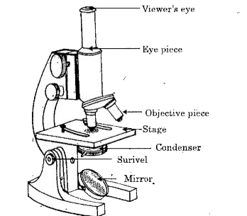
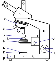
Label Microscope Diagram - EnchantedLearning.com Label Microscope Diagram Using the terms listed below, label the microscope diagram. Inventions and Inventors arm - this attaches the eyepiece and body tube to the base. base - this supports the microscope. body tube - the tube that supports the eyepiece. coarse focus adjustment - a knob that makes large adjustments to the focus.
Adipose Tissue Under Microscope with Labeled Diagram Adipose tissue under microscope labeled diagram. Now, I will show you the 2 types of adipocytes (white and brown) and adipose tissue with the labeled diagram. This might help you understand the basic structure and identify the adipose tissue under a microscope. First, let's see the labeled diagram of white and brown adipocytes.
Parts of the Microscope (Labeled Diagrams) Simple microscope labelled diagram Image created with Biorender Tube/Body Tube It serves as the connector between the eyepiece/ocular and objective lenses. Objective lenses The lenses have varying magnifying power, which typically consists of 10x, 40x, and 100x.
Microscope Parts, Types & Diagram | What is a Microscope? Microscope Diagram There are many illustrations available for the diagram of a light microscope. The essential parts include the head, base, arms, lenses, and lights. In diagrams, one...
Parts of the Microscope with Labeling (also Free Printouts) A microscope is one of the invaluable tools in the laboratory setting. It is used to observe things that cannot be seen by the naked eye. Table of Contents 1. Eyepiece 2. Body tube/Head 3. Turret/Nose piece 4. Objective lenses 5. Knobs (fine and coarse) 6. Stage and stage clips 7. Aperture 9. Condenser 10. Condenser focus knob 11. Iris diaphragm


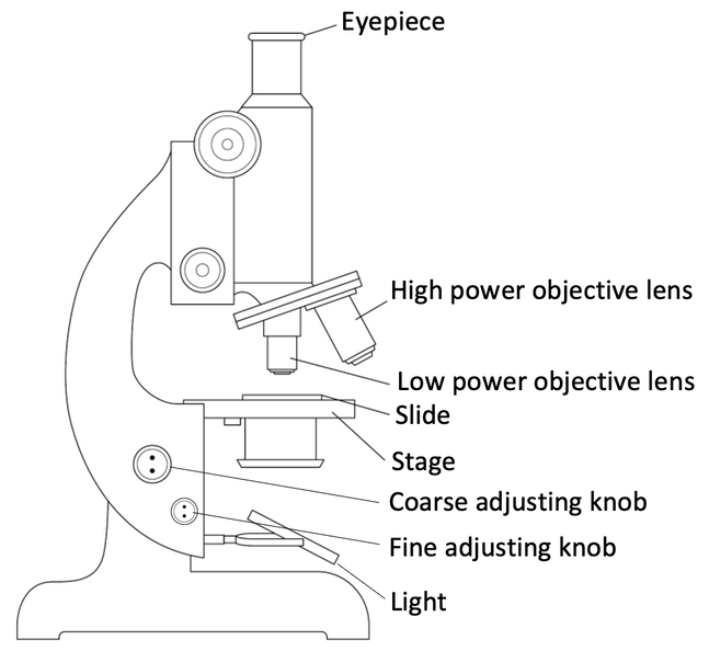


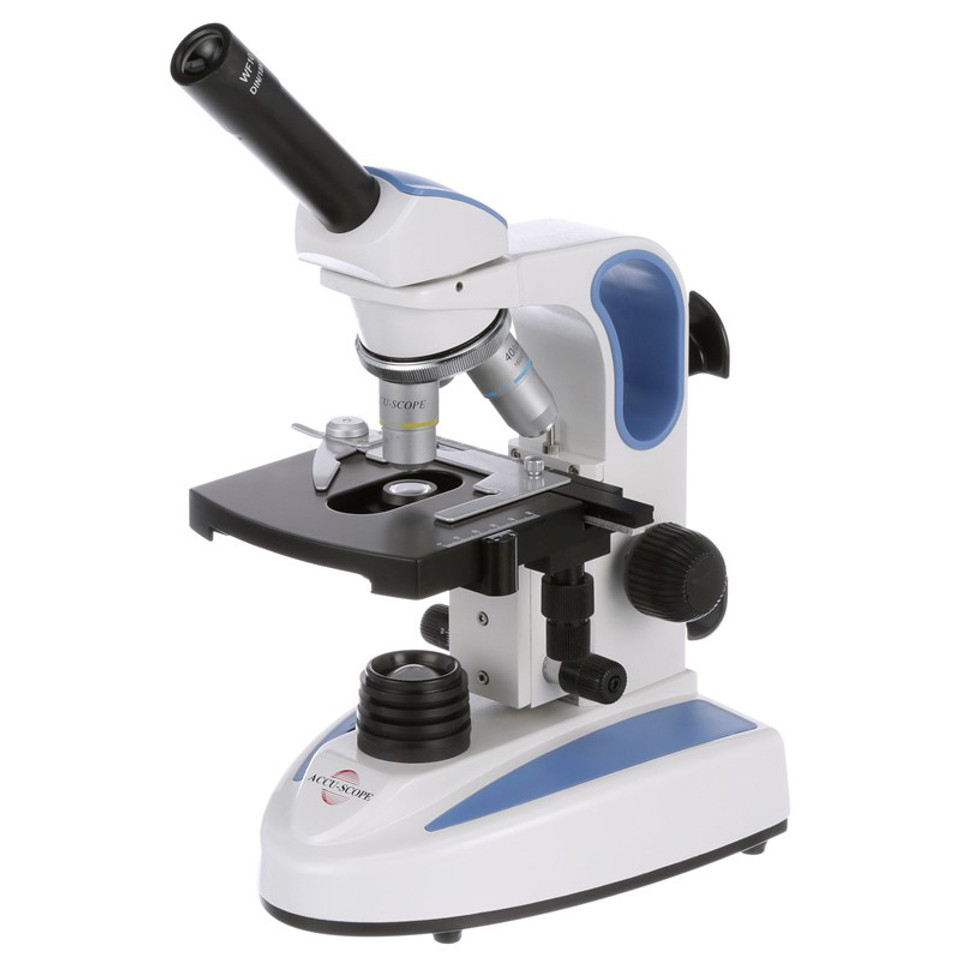










Post a Comment for "41 microscope diagram with labels"