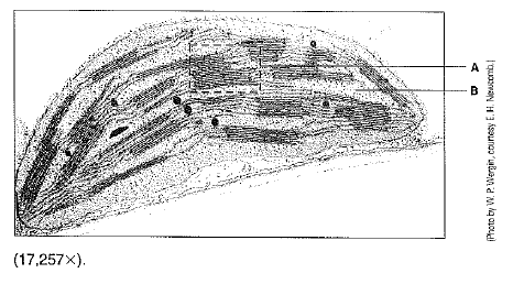
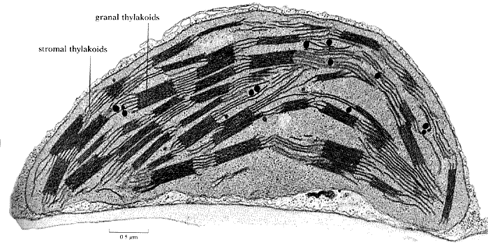
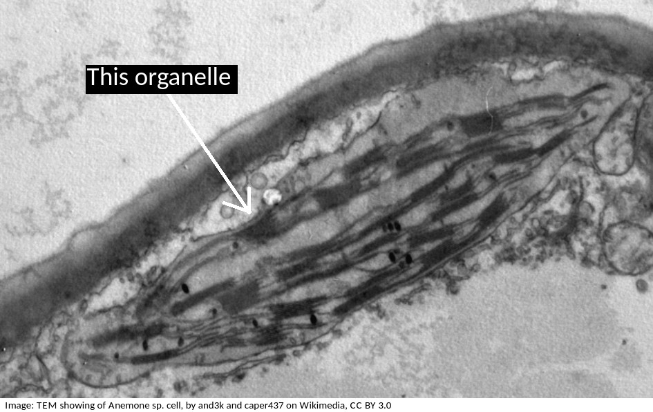
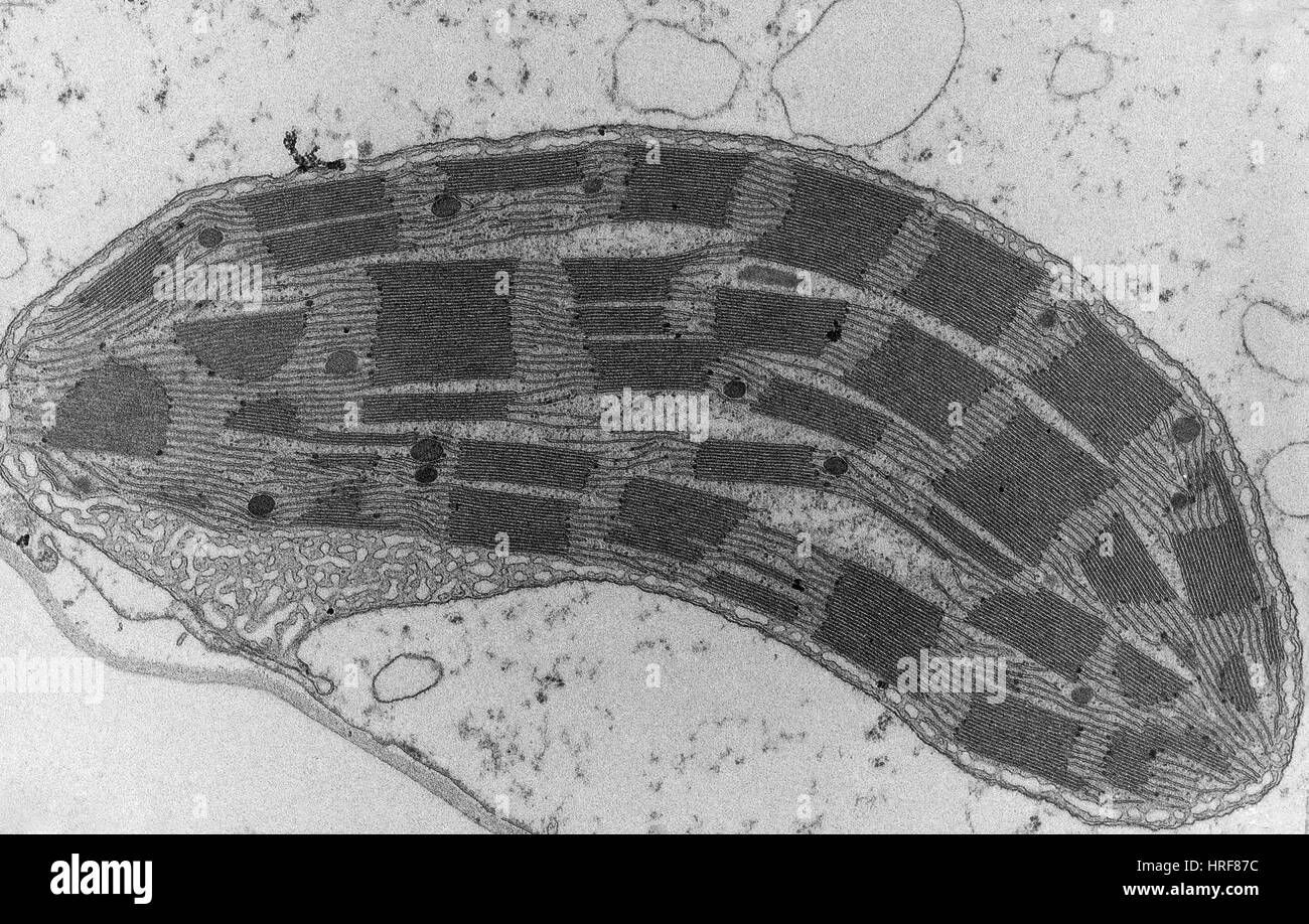
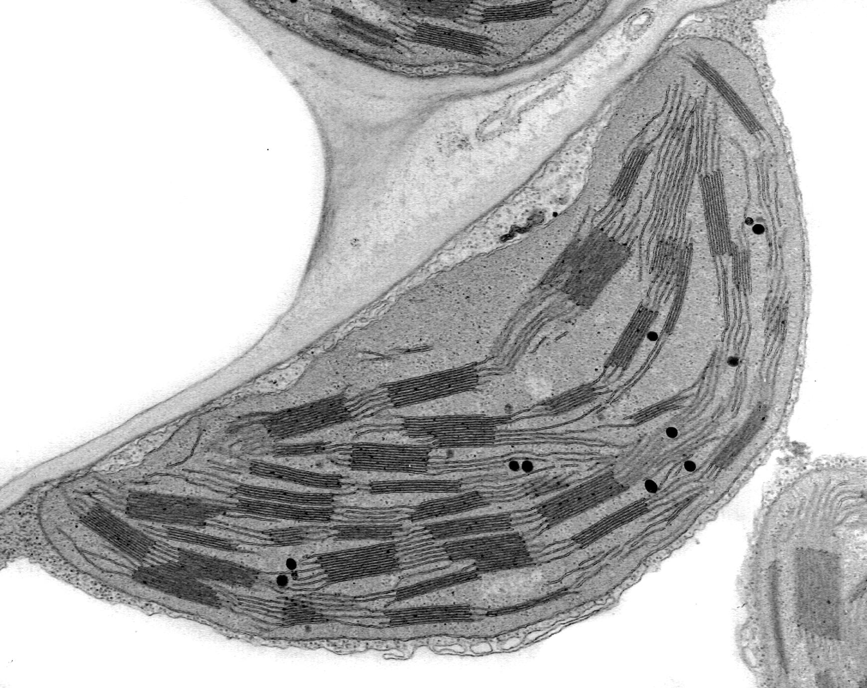
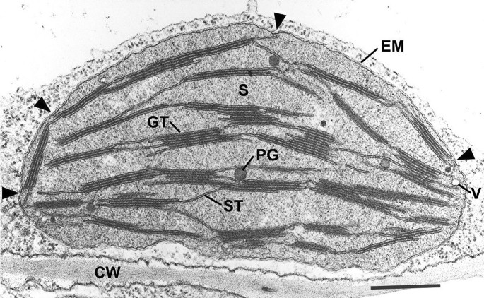
42 label the transmission electron microscope image of a chloroplast below
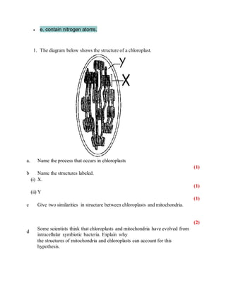
Labeling the Cell Flashcards | Quizlet Flashcards Learn Test Match Created by mersadies_morgan Teacher Terms in this set (27) Label the structures of the plasma membrane and cytoskeleton. Label the membranous organelles. Label these nuclear structures and ribosomes. Label the types of plasma membrane lipids. Label the types of plasma membrane proteins. Label the outer leaflet. Cells and magnification Flashcards | Quizlet 9) The figure below shows a photograph of a chloroplast taken with an electron microscope a- Name the parts of the chloroplast labelled A and B (2 marks).
Microscopy: Intro to microscopes & how they work (article ... - Khan ... In transmission electron microscopy ( TEM ), in contrast, the sample is cut into extremely thin slices (for instance, using a diamond cutting edge) before imaging, and the electron beam passes through the slice rather than skimming over its surface ^5 5. TEM is often used to obtain detailed images of the internal structures of cells.

Label the transmission electron microscope image of a chloroplast below
DP Biology: Ultrastructure of cells quiz 1.2 - Thinkib.net The electron microscope image below shows an organelle found in eukaryote cells. What is the name of the organelle? Chloroplast. Mitochondrion. Electron microscopes - Cell structure - Edexcel - BBC Bitesize the scanning electron microscope (SEM) has a large depth of field. so can be used to examine the surface structure of specimens TEMs have a maximum magnification of around ×1,000,000, but images ... Transmission electron micrograph of a chloroplast - Getty Images Creative #: 1131215162 License type: Royalty-free Collection: Image Source Location: United States Release info: No release required Similar images View all Plant Biological Cell Chloroplast Electron Microscope Chlorophyll Photosynthesis Begonia Biological Process Botany Close-up Color Enhanced Color Image Cross Section Electron Micrograph
Label the transmission electron microscope image of a chloroplast below. Click here for - WordPress.com The diagram shows a chloroplast as seen with an electron microscope. ... The figure below shows a photograph of a chloroplast taken with an electron ... Bio 150 - Microscopy and Cell Structure (Lab 5) Flashcards A microscope that has a large working distance that is useful for viewing and manipulating larger specimens but only magnifies the specimen to a limited extent. Electron Microscope Microscopes that use a beam of electrons to bounce off the specimen and a very sensitive detector to resolve cellular structures at incredibly high magnification. Electron Microscopy Images - Dartmouth Sep 10, 2021 ... We have a library of images recorded using our scanning and transmission electron microscopes. Some are shown below and others elsewhere. AP Biology Study the electron micrographs on page 110 of your text. ... Now label the diagram of the chloroplast below being to include the outer membrane, ...
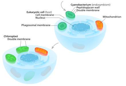
photosynthesis.pdf - 228 — Chloroplasts 1. Label the transmission ... View photosynthesis.pdf from BIO 123 at Üsküdar American Academy High School. 228 — Chloroplasts 1. Label the transmission electron microscope image of a chloroplast below: a) stroma b) stroma Electron Microscopy Views of Dimorphic Chloroplasts in C4 Plants Chloroplasts in C4 plants exhibit structural dimorphism because thylakoid architectures vary depending on energy requirements. Advances in electron microscopy imaging capacity and sample preparation technologies allowed characterization of thylakoid structures and their macromolecular arrangements with unprecedented precision mostly in C3 plants. 10.1: Plant Cell Structure and Components - Biology LibreTexts Figure 10.1. 12: This image shows the same Elodea leaf cells again, this time with the cell wall, cell membrane, and tonoplast of one of the cells labeled. The cell walls are visible as thicker lines between the cells. The plasma membrane and tonoplast locations must be inferred. Mitochondria and chloroplasts (article) | Khan Academy Mitochondria are the "powerhouses" of the cell, breaking down fuel molecules and capturing energy in cellular respiration. Chloroplasts are found in plants and algae. They're responsible for capturing light energy to make sugars in photosynthesis. Mitochondria and chloroplasts likely began as bacteria that were engulfed by larger cells (the ...
What are the labels of the transmission electronic microscope image ... What are the labels of the transmission electronic microscope image of a chloroplast 1 See answer Advertisement ireenys3004 Answer: Explanation: Transfer RNA (tRNA) precursors undergo endoribonucleolytic processing of their 5' and 3' ends. 5' cleavage of the precursor transcript is performed by ribonuclease P (RNase P). Transmission electron microscope images of chloroplasts in WT (a, b ... Transmission electron microscope images of chloroplasts in WT (a, b) and wsl9 mutant (c, d) seedlings. Scale bar, 0.25 um in a, c; 0.15 um in (b, d) Source publication +4 WSL9 Encodes an... Chloroplast | Definition, Function, Structure, Location, & Diagram Chloroplasts are roughly 1-2 μm (1 μm = 0.001 mm) thick and 5-7 μm in diameter. They are enclosed in a chloroplast envelope, which consists of a double membrane with outer and inner layers, between which is a gap called the intermembrane space. Biology paper 1 questions Flashcards | Quizlet The image shows a phagocytic white blood cell as seen with a transmission electron microscope. Which features can be found both within this cell and in a ...
Electron Microscopy Views of Dimorphic Chloroplasts in C4 Plants As reviewed by Staehelin in 2003, the study of chloroplast structure has advanced with enhancements in light and electron microscopy from two-dimensional transmission electron microscopy (TEM) imaging to three-dimensional (3D) electron tomography (ET) and with improvements in sample preservation from chemically preservation to freeze-fracture to...
Solved Examine the transmission electron microscope image - Chegg Question: Examine the transmission electron microscope image below. a) Identify ... a) Here A= Endoplasmic Reticulum B= Chloroplast C= Cytoplasm D= Golgi ...
I ntroduction to Photosynthesis - images organetles catted chloroplasts inside ... electrons that provide the energy ... label the transmission electron micrograph (TEM) of a chloroplast below:.
label the transmission electron microscope image of a chloroplast below ... label the transmission electron microscope image of a chloroplast below Electron biogenesis photosystem localized translation If you are searching about Transmission electron micrograph of animal cell - Stock Image - G450 you've came to the right web.
The Transmission Electron Microscope | CCBER - UC Santa Barbara Transmission electron microscopes (TEM) are microscopes that use a particle beam of electrons to visualize specimens and generate a highly-magnified image. TEMs can magnify objects up to 2 million times. In order to get a better idea of just how small that is, think of how small a cell is. It is no wonder TEMs have become so valuable within the ...
Classical transmission electron microscopy (TEM) led to the formulation ... Classical transmission electron microscopy (TEM) led to the formulation of the helical model of thylakoid architecture In this section we review the progression of TEM observations that led to the formulation of the generally accepted helical model of granum structure.
Transmission electron microscopic images of chloroplasts and... Transmission electron microscopic images of chloroplasts and mitochondria in 15-day-old leaves from PRORP1 RNAi mutants and wild-type plants. (A, B) Ultrastructure of chloroplasts and...
Correlative light electron microscopy using gold nanoparticles as ... Correlative light electron microscopy (CLEM) is a powerful tool in bioimaging, as it combines the ability to image living cells over large fields of view with molecular specificity using light ...
Mitochondria and Chloroplasts - Principles of Biology Figure 1 This transmission electron micrograph shows a mitochondrion as viewed with an electron microscope. Notice the inner and outer membranes, the cristae, and the mitochondrial matrix. (credit: modification of work by Matthew Britton; scale-bar data from Matt Russell) Like mitochondria, chloroplasts also have their own DNA and ribosomes.
Transmission electron micrograph of a chloroplast - Getty Images Creative #: 1131215162 License type: Royalty-free Collection: Image Source Location: United States Release info: No release required Similar images View all Plant Biological Cell Chloroplast Electron Microscope Chlorophyll Photosynthesis Begonia Biological Process Botany Close-up Color Enhanced Color Image Cross Section Electron Micrograph
Electron microscopes - Cell structure - Edexcel - BBC Bitesize the scanning electron microscope (SEM) has a large depth of field. so can be used to examine the surface structure of specimens TEMs have a maximum magnification of around ×1,000,000, but images ...
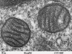
DP Biology: Ultrastructure of cells quiz 1.2 - Thinkib.net The electron microscope image below shows an organelle found in eukaryote cells. What is the name of the organelle? Chloroplast. Mitochondrion.











Post a Comment for "42 label the transmission electron microscope image of a chloroplast below"