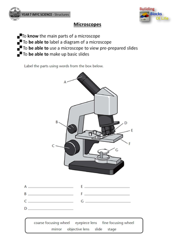
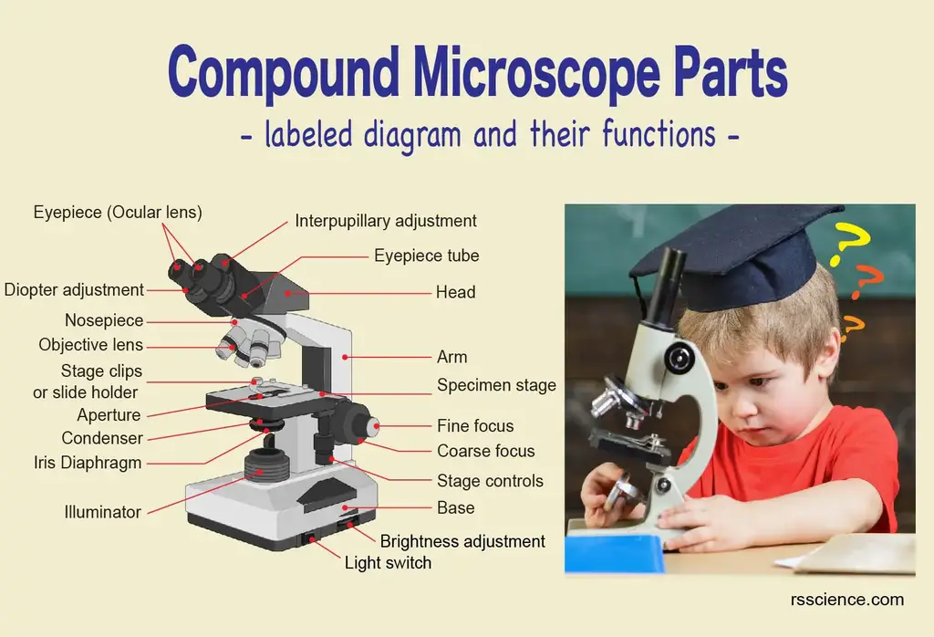
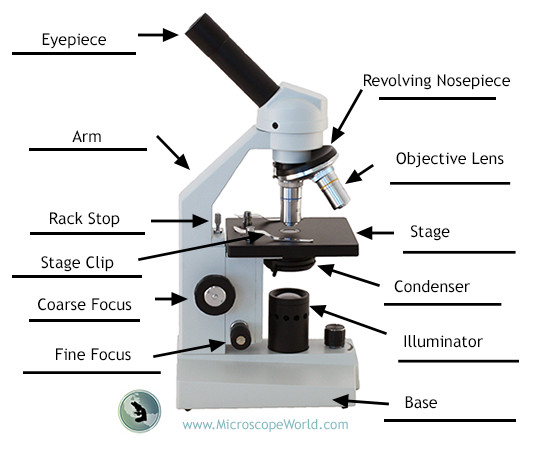
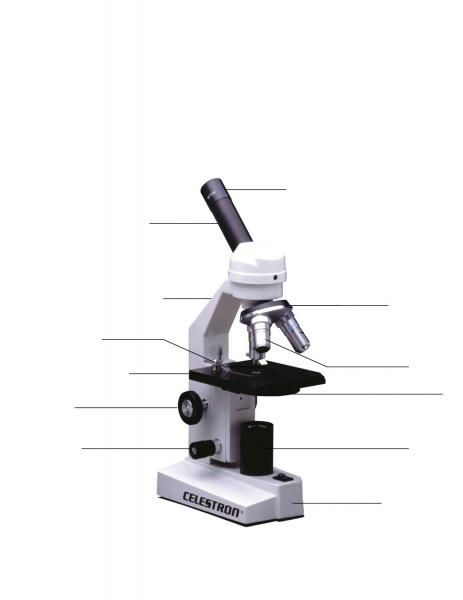
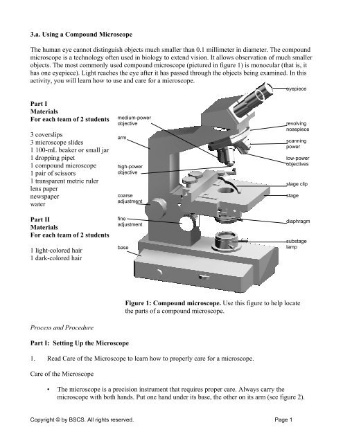
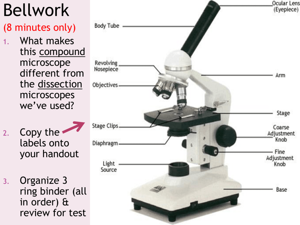
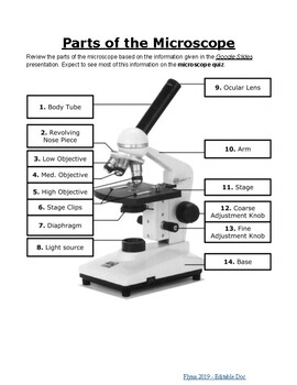
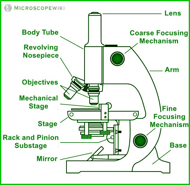
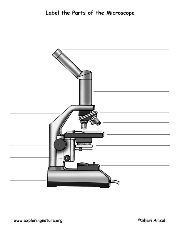
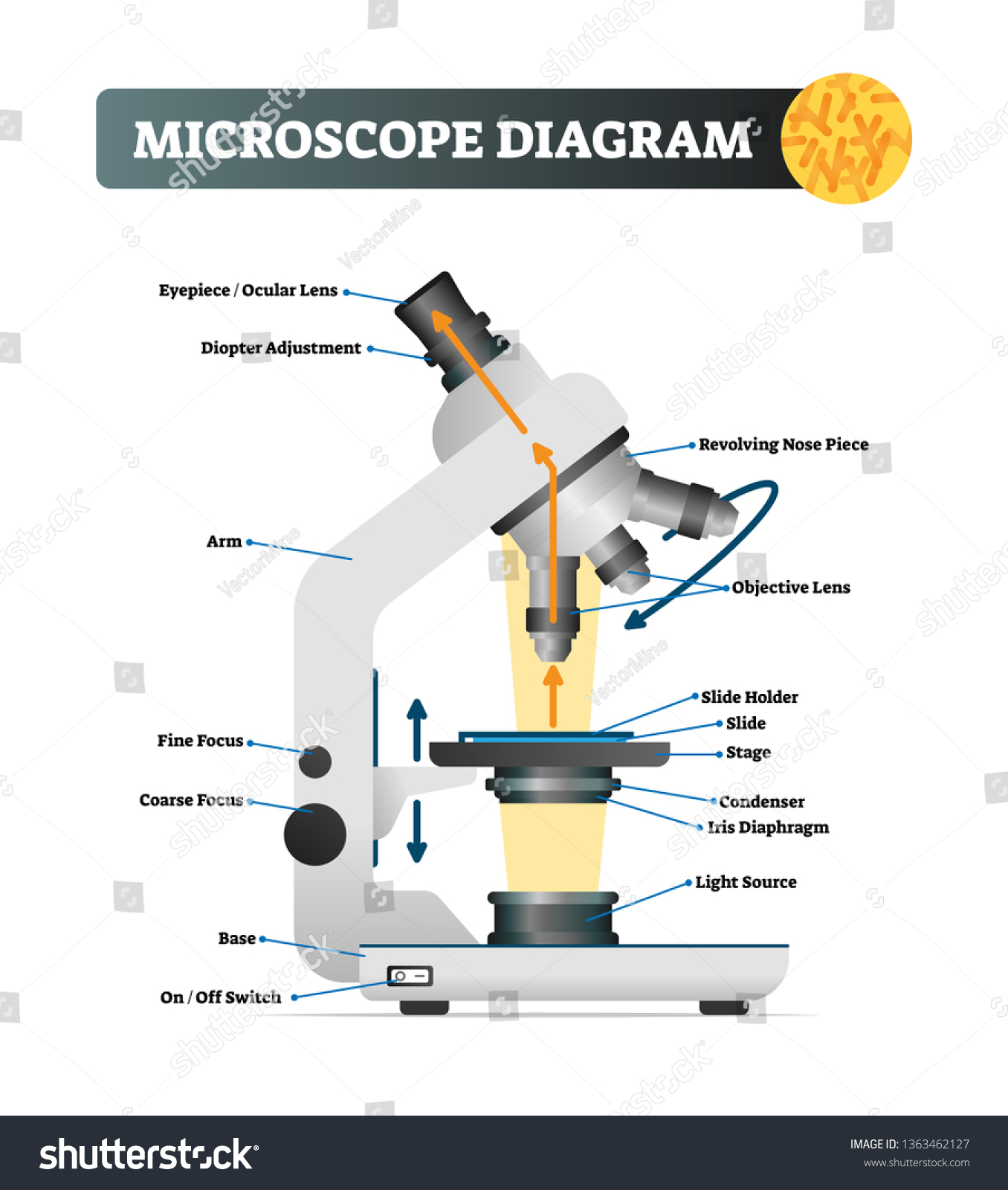
41 microscope picture with labels
histology.leeds.ac.ukHome: The Histology Guide You can see histological slides on the pages and can turn labels on or off to help them identify features. In some cases, there is a section like a 'virtual microscope' - you can scan around a large picture using the mouse and try to identify features. This emulates as closely as possible the experience of using a microscope. Home: The Histology Guide You can see histological slides on the pages and can turn labels on or off to help them identify features. In some cases, there is a section like a 'virtual microscope' - you can scan around a large picture using the mouse and try to identify features. This emulates as closely as possible the experience of using a microscope. We've also recently introduced a new feature, where …
Alcon Inc. (ALC) CEO David Endicott on Q2 2022 Results - Earnings Call ... Core diluted earnings per share in the second quarter of 2022 were $0.63, up from $0.56 last year. Before I discuss our outlook for the remainder of 2022, I'll touch on a couple of cash flow and ...
Microscope picture with labels
Amazon: Champion clothing up to 69% off, Hanes women's shorts only $9 ... Amazon has some great buys right now including Champion clothing for adults and kids on sale up to 69% off, Wireless Bluetooth Earbuds Set with Charging Case for $16.97 (reg. $29.99), school ... Microarray Technology - Genome.gov Microarray technology is a general laboratory approach that involves binding an array of thousands to millions of known nucleic acid fragments to a solid surface, referred to as a "chip.". The chip is then bathed with DNA or RNA isolated from a study sample (such as cells or tissue). Complementary base pairing between the sample and the ... Hyperglycemia promotes myocardial dysfunction via the ERS-MAPK10 ... Pulse-wave Doppler images of mitral inflow from the apical 4 ... Beijing, China), and rhodamine-labeled wheat germ agglutinin (WGA) (1.25 mg/mL; ZD0510, Vector Laboratory, Burlingame, CA, USA ...
Microscope picture with labels. KEYENCE TV : VR-3000 Series | KEYENCE America The VR-3000 Wide-Area 3D Measurement System combines the advanced optical design of our digital microscopes with the high-speed, high-accuracy measurement and 3D technology of our displacement gauges to complete instant 3D measurements. This new technology captures 3D data over a wide area with just a click of a button, eliminates measurement ... Hypodermis of the Skin Anatomy and Physiology - Verywell Health The hypodermis is the innermost layer of the skin located under the dermis (outer layer) and the epidermis (middle layer). The thickness of the hypodermis varies in different regions of the body and can vary considerably between different people. The hypodermis layer also provides shaping and contouring. For those assigned male at birth, the ... ZEISS Axioscan 7 Microscope Slide Scanner Digitize your specimens with Axioscan 7 – the reliable, reproducible way to create high-quality virtual microscope slides. Axioscan 7 combines qualities that you would not expect to get in a slide scanner: high speed digitization and outstanding image quality plus an unrivaled variety of imaging modes are all available in a fully automated and easy to operate system. Motility Test (Theory) - Amrita Vishwa Vidyapeetham Virtual Lab Three methods are employed for motility determination depending on the pathogenic capability of the organisms. For nonpathogens, there are two slide techniques that one might use. For pathogens, tube method can be used. I) Slide methods for non-pathogens include 1. Wet Mount slide 2. Hanging Drop slide 1. Wet Mount slide
Cell Size and Scale - University of Utah Smaller cells are easily visible under a light microscope. It's even possible to make out structures within the cell, such as the nucleus, mitochondria and chloroplasts. Light microscopes use a system of lenses to magnify an image. The power of a light microscope is limited by the wavelength of visible light, which is about 500 nm. The most powerful light microscopes can … Ti2-LAPP | Photostimulation & TIRF - Nikon Instruments Inc. An in vitro preparation of fluorescently-labeled microtubules (tetramethylrhodamine and Alexa 647) and tubulin binding proteins (Alexa 488) was imaged in three different wavelengths using the H-TIRF illuminator and the gradation ND filter. Incident angles can be automatically adjusted for multiple wavelengths. Negative Staining- Principle, Reagents, Procedure and Result Procedure of Negative Staining 1. Place a very small drop (more than a loop full, less than a free falling drop from the dropper) of nigrosin near one end of a well-cleaned and flamed slide. 2. Remove a small amount of the culture from the slant with an inoculating loop and disperse it in the drop of stain without spreading the drop. 3. Looking at the Structure of Cells in the Microscope A typical animal cell is 10–20 μm in diameter, which is about one-fifth the size of the smallest particle visible to the naked eye. It was not until good light microscopes became available in the early part of the nineteenth century that all plant and animal tissues were discovered to be aggregates of individual cells. This discovery, proposed as the cell doctrine by Schleiden and …
Anatomy Chart - How to Make Medical Drawings and Illustrations … Histology studies microscopic anatomy such as tissues and cells visible only under a microscope. Anatomy charts serve two main purposes: education in the form of anatomy worksheets and presentation in the form of simple healthcare illustrations. Anatomy illustrations are pre-made illustrations with descriptions of a particular part, or system of the body. Anatomy … › set › classroom-objectsClassroom Objects – ESL Flashcards Description. Here are some flashcards for teaching the names of common school supplies and objects usually found in classrooms. The set includes both items that students (should) have in their own school bags as well as items likely to be found on the teacher’s desk only, such as a stapler. IFN-γ stimulated murine and human neurons mount anti-parasitic defenses ... D Schematic of host cells labeled by RCre versus GCre parasites. ... The stained cultures were analyzed by confocal microscope. A Representative images of stained cultures from wild type (WT) mice ... Super-Resolution Microscope System - Nikon Instruments Inc. This larger imaging area enables very high throughput for applications/samples that benefit from larger fields of view, such as neurons, reducing the amount of time and effort required to obtain data. Reconstructed image size: 1024 x 1024 pixels (33 μm x 33 μm with a 100X objective)
What is Electron Microscopy? - UMASS Medical School Because the size of the raster at the specimen is much smaller than the viewing screen of the CRT, the final picture is a magnified image of the specimen. Appropriately equipped SEMs (with secondary, backscatter and X-ray detectors) can be used to study the topography and atomic composition of specimens, and also, for example, the surface distribution of immuno-labels.
Light-driven single-cell rotational adhesion frequency assay Our scRAFA exploits a microfluidic platform integrated with versatile optothermal manipulation and optical imaging to trap and rotate single cells while monitoring the sequential cell rotation and cell-substrate adhesion [19, 20] (Fig. 2a).As a demonstration, Figs. 2b, c show the successive images of light-driven trapping and rotation of a single S. cerevisiae above a substrate (Additional ...
Super-Resolution Microscope System - Nikon Corporation Healthcare ... Two-color TIRF-SIM imaging of growth cone of NG108 cell labeled with Alexa Fluor ® 488 for F-actin (green) and Alexa Fluor ® 555 for microtubules (orange) Reconstructed image size: 2048 x 2048 pixels (66 μm x 66 μm with a 100X objective)
Counting of Fluorescent Particles - Amrita Vishwa Vidyapeetham Fluorescently labeled cells under a fluorescent microscopy In biomedical research, Fluorescent assay will allow researchers to identify sub cellular structures such as proteins, chromosomes, genes and mutation in genes which further helps for its quantification.
All Colorado judges recommended for retention in 2022 However, the results of Colorado's retention process do not provide the complete picture of judicial performance. A total of 164 judges were eligible for retention in 2022, but only 140 received evaluations and 135 chose to remain on the ballot. Judges may opt to resign or retire prior to their retention for multiple reasons, including the expectation of a negative performance evaluation.
Protozoa Cells Under Microscope - central nervous system protozoal ... Protozoa Cells Under Microscope - 8 images - amoeba under microscope 400x labeled micropedia, Menu ≡ ╳ Home ; Login & Register ; Contact ; Home; Protozoa Cells Under Microscope; Protozoa Cells Under Microscope. Published by Willie; Saturday, August 13, 2022 ...
Fluorescence In Situ Hybridization (FISH) - Genome.gov The fluorescently labeled DNA finds its matching segment on one of the chromosomes, where it sticks. By looking at the chromosomes under a microscope, a researcher can find the region where the DNA is bound because of the fluorescent dye attached to it. This information thus reveals the location of that piece of DNA in the starting genome.
Light Microscope (Assignment) - Amrita Vishwa Vidyapeetham To illustrate the working of a microscope, follow the instructions in the simulator. Students Assignment . Are there any other objective used in light microscope other than 4X, 10X, 40Xand 100X? If YES, specify them? Consider a light microscope without iris diaphragm. How the images at 4 X, 10 X, 40 X and 100 X differ?
Metaphase - Genome.gov Metaphase is a stage during the process of cell division (mitosis or meiosis). Normally, individual chromosomes are spread out in the cell nucleus. During metaphase, the nucleus dissolves and the cell's chromosomes condense and move together, aligning in the center of the dividing cell. At this stage, the chromosomes are distinguishable when ...
› confocal-microscopes › lsm-900LSM 900 with Airyscan 2 – Compact Confocal Microscope for ... In microscopy, this translates into the best contrast and resolution while maintaining minimum light exposure. LSM 900, your compact confocal microscope, provides this with components optimized to deliver the best imaging results. Get high-end confocal imaging in a small footprint. Improve any confocal experiment with LSM Plus.
Parasite reliance on its host gut microbiota for nutrition and survival ... D BODIPY and DAPI labeled images of fat body cells from 3LL GN D. melanogaster larvae inoculated with A. pomorum only, A. pomorum and Bacillus. sp., or A. pomorum, Bacillus. sp. and Rhodococcus sp ...
LSM 900 with Airyscan 2 – Compact Confocal Microscope for In microscopy, this translates into the best contrast and resolution while maintaining minimum light exposure. LSM 900, your compact confocal microscope, provides this with components optimized to deliver the best imaging results. Get high-end confocal imaging in a small footprint. Improve any confocal experiment with LSM Plus.
› microscopy › intZEISS Axioscan 7 Microscope Slide Scanner Digitize your specimens with Axioscan 7 – the reliable, reproducible way to create high-quality virtual microscope slides. Axioscan 7 combines qualities that you would not expect to get in a slide scanner: high speed digitization and outstanding image quality plus an unrivaled variety of imaging modes are all available in a fully automated and easy to operate system.
Microscope Diagram - cell division of e coli with continuous media flow ... Microscope Diagram - 15 images - give a well labelled diagram of compound microscope using of typical, bio tem biological transmission electron microscope university, labelled microscope diagram gcse micropedia, a compound microscope diagram micropedia,
› cemf › whatisemWhat is Electron Microscopy? - UMASS Medical School Conventional scanning electron microscopy depends on the emission of secondary electrons from the surface of a specimen. Because of its great depth of focus, a scanning electron microscope is the EM analog of a stereo light microscope. It provides detailed images of the surfaces of cells and whole organisms that are not possible by TEM.
› crazy-random-stuff › microscopeMicroscope For Sale in Clondalkin, Dublin from Lndnll Microscope, Used Crazy Random ... Personalised ties with any picture and any text you like. €15.00 ... Custom Printed Stickers / Decals / Wedding Labels. €25.00
Super-Resolution Microscope System - Nikon Europe B.V. Multi-color super-resolution imaging can be carried out using both activator-reporter pairs for sequential activation imaging and activator-free labels for continuous activation imaging. This flexibility allows users to easily gain critical insights into the localization and interaction properties of multiple proteins at the molecular level.
Pyramidal neuron subtype diversity governs microglia states in the ... Fig. 1: PN subtypes locally control microglia density in the cortex. a, Schematic of control and Fezf2 -KO S1 cortex. PN classes are colour coded. b, Representative micrograph of P7 control and...
Micro Module 1 Flashcards | Quizlet Study with Quizlet and memorize flashcards containing terms like Move the terms into the correct empty boxes to complete the concept map., Drag the images and/or statements to their corresponding class to test your understanding of the main types of microbes., Drag the images or descriptions to their corresponding class to test your understanding of the cellular organization …
Motility Test (Procedure) - Amrita Vishwa Vidyapeetham Label the slide with the name of the organism Place 15 - 20 uL of the culture in the middle of the slide Lower a clean cover slip over the drop as though it were hinged at one side avoiding bubbles Examine the preparation under microscope first under 4 x followed by 40 x and 100x magnification Identify the motile organisms
Motility Test - Principle, Procedure, Uses and Interpretation Method. Touch a straight needle to a colony of a young (18- to 24-hour) culture growing on agar medium. Stab once to a depth of only 1/3 to ½ inch in the middle of the tube. Be sure to keep the needle in the same line it entered as it is removed from the medium. Incubate at 35°-37°C and examine daily for up to 7 days.
Gram Staining: Principle, Procedure, Interpretation, Examples and Animation Procedure of Gram Staining. Take a clean, grease free slide. Prepare the smear of suspension on the clean slide with a loopful of sample. Crystal Violet was poured and kept for about 30 seconds to 1 minutes and rinse with water. Flood the gram's iodine for 1 minute and wash with water. Then ,wash with 95% alcohol or acetone for about 10-20 ...
Classroom Objects – ESL Flashcards Description. Here are some flashcards for teaching the names of common school supplies and objects usually found in classrooms. The set includes both items that students (should) have in their own school bags as well as items likely to be found …
ECLIPSE Ti2 Series | Inverted Microscopes - Nikon Instruments Inc. Volume Contrast technique utilizes a series of label-free, brightfield images captured at various Z-depths to assemble a phase distribution image. Volume Contrast renders cells easily identifiable as objects for automated counting and area analysis.
Microscope For Sale in Clondalkin, Dublin from Lndnll - Adverts.ie Microscope. Click here to send this ad to a friend Microscope . Asking price: €40 . Make an offer: ... Personalised ties with any picture and any text you like. €15.00 . Personalised Rectangular Metal Photo Keyring. €10.00 . Personalised Flipflops. €25.00 . NEW 40KG Mini Electronic Digital LCD Hand Hanging Luggage Kitchen Weight Hook Scale. €18.90 . …
› books › NBK26880Looking at the Structure of Cells in the Microscope ... The phase-contrast microscope and, in a more complex way, the differential-interference-contrast microscope, exploit the interference effects produced when these two sets of waves recombine, thereby creating an image of the cell's structure . Both types of light microscopy are widely used to visualize living cells.
Light Microscope (Theory) - Amrita Vishwa Vidyapeetham Microscope Microscope is an optical instrument that uses lens or combination of lens to produce magnified images that are too small to seen by unaided eye. Microscope provides the enlarged view that helps in examining and analyzing the image.
Hyperglycemia promotes myocardial dysfunction via the ERS-MAPK10 ... Pulse-wave Doppler images of mitral inflow from the apical 4 ... Beijing, China), and rhodamine-labeled wheat germ agglutinin (WGA) (1.25 mg/mL; ZD0510, Vector Laboratory, Burlingame, CA, USA ...
Microarray Technology - Genome.gov Microarray technology is a general laboratory approach that involves binding an array of thousands to millions of known nucleic acid fragments to a solid surface, referred to as a "chip.". The chip is then bathed with DNA or RNA isolated from a study sample (such as cells or tissue). Complementary base pairing between the sample and the ...
Amazon: Champion clothing up to 69% off, Hanes women's shorts only $9 ... Amazon has some great buys right now including Champion clothing for adults and kids on sale up to 69% off, Wireless Bluetooth Earbuds Set with Charging Case for $16.97 (reg. $29.99), school ...













Post a Comment for "41 microscope picture with labels"